
B27-like Animal Origin-Fre Supplement
NEW
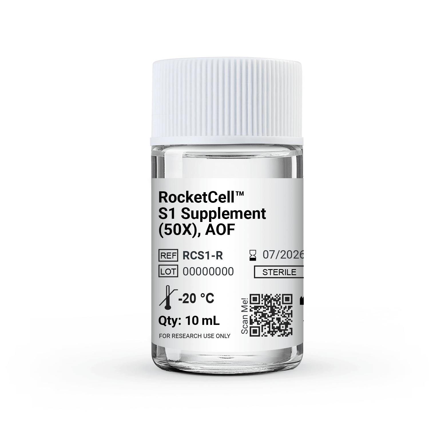
RocketCell™ S1 Supplement (50X), AOF
B27-like, Animal Origin-Free Supplement
RocketCell™ S1 Supplement (50X), AOF

Comprehensive Antioxidant protection
An optimized blend of antioxidant enzymes and vitamins that helps protect sensitive cells from oxidative damage, supporting high viability even at low plating densities.

3D-Validated Specifically optimized for use within VitroGel® hydrogel systems for spheroid and organoid applications. While optimized for VitroGel® hydrogels, it also supports animal-based ECMs cultures for spheroids, neuronal cultures, complex co-cultures, and organoid model development.

Drop-in replacement
Protocol-compatible with standard B27 workflows at equivalent working concentrations.

Retinoid-Competent Formulation
Supports baseline CNS neuronal maintenance, maturation, and long-term culture stability.
Broad and Versatile
Supports organoid, tumoroid, hippocampal and cortical neuron cultures, iPSC-derived neuron maturation, cancer stem cell, and cardiomyocyte maintenance.
RocketCell™ S1 Supplement, AOF is an animal origin-free replacement for conventional B27 supplements, engineered for the long-term survival, maturation, and functional maintenance of CNS neurons and a broad spectrum of other cell types. All components are of recombinant, synthetic origin, eliminating the batch-to-batch variability and safety concerns inherent in animal-derived additives.
This is the all-purpose formulation — the right choice when the primary goal is maximizing long-term cell viability without directing cell fate. Supplied as a 50X liquid concentrate for use with standard neurobasal or serum-free basal media.
Challenges We Solve


Common Challenges

Lack of a reliable animal origin-free formulation to support GMP process.

Variability in culture conditions across experiments.

Unwanted signaling that can interfere with targeted differentiation.

Difficulty in maintaining long-term cultures
How RocketCell™ S1 Supplement, AOF Solves The Issues
Animal origin-free formulation for GMP transition
B27- like AOF formulation that simply transition to scalable and controlled GMP workflows.

Standardized B27-like formulation
Provides a standardized formulation to support reproducible culture conditions across various tissue sources and cell models.
Formulations for controlled differentiation
Available in minus Insulin and minus Vitamin A formulations for greater control over differentiation protocols.

Sustained long-term cell survival
Supports cell viability during extended culture of advanced 3D models. Supports the maturation and maintenance of differentiated cell models, including iPSC-derived neurons and cardiomyocytes.
Protocols and Resources
 Protocol

Data and References
ORGANOIDS
Cryo-Recovery and Re-Culturing Intestinal Organoid with RocketCell™ S1 (B27-like) & S2 (N2-like) Supplements, AOF
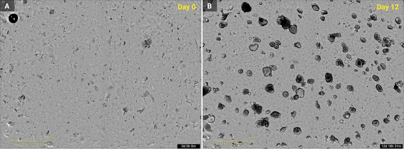
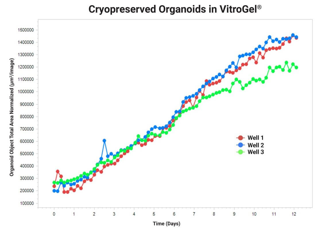
Cryo-recovery and re-culturing of intestinal organoids using RocketCell™ S1 (B27-like) & S2 (N2-like) Supplements, AOF.
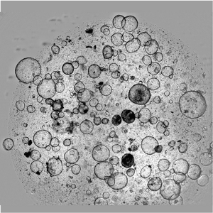
Representative bright-field images show cryopreserved intestinal organoids following recovery at Day 0 (A) and subsequent growth at Day 12 (B) in VitroGel®. Quantitative analysis demonstrates progressive organoid expansion over 12 days across three replicate wells, indicating successful recovery and sustained growth following cryopreservation.
Mouse Intestinal Organoid Cultured using RocketCell™ S1 (B27-like) & S2 (N2-like) Supplements, AOF
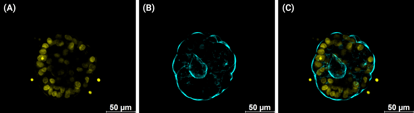
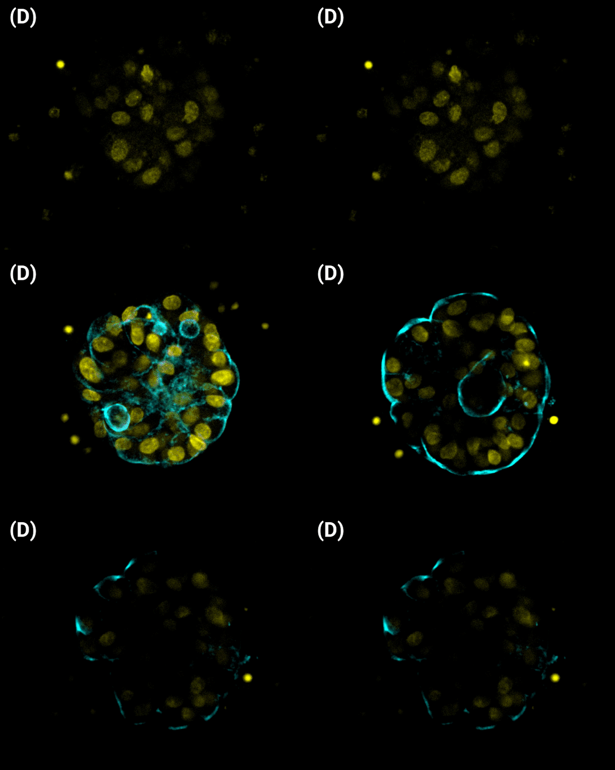
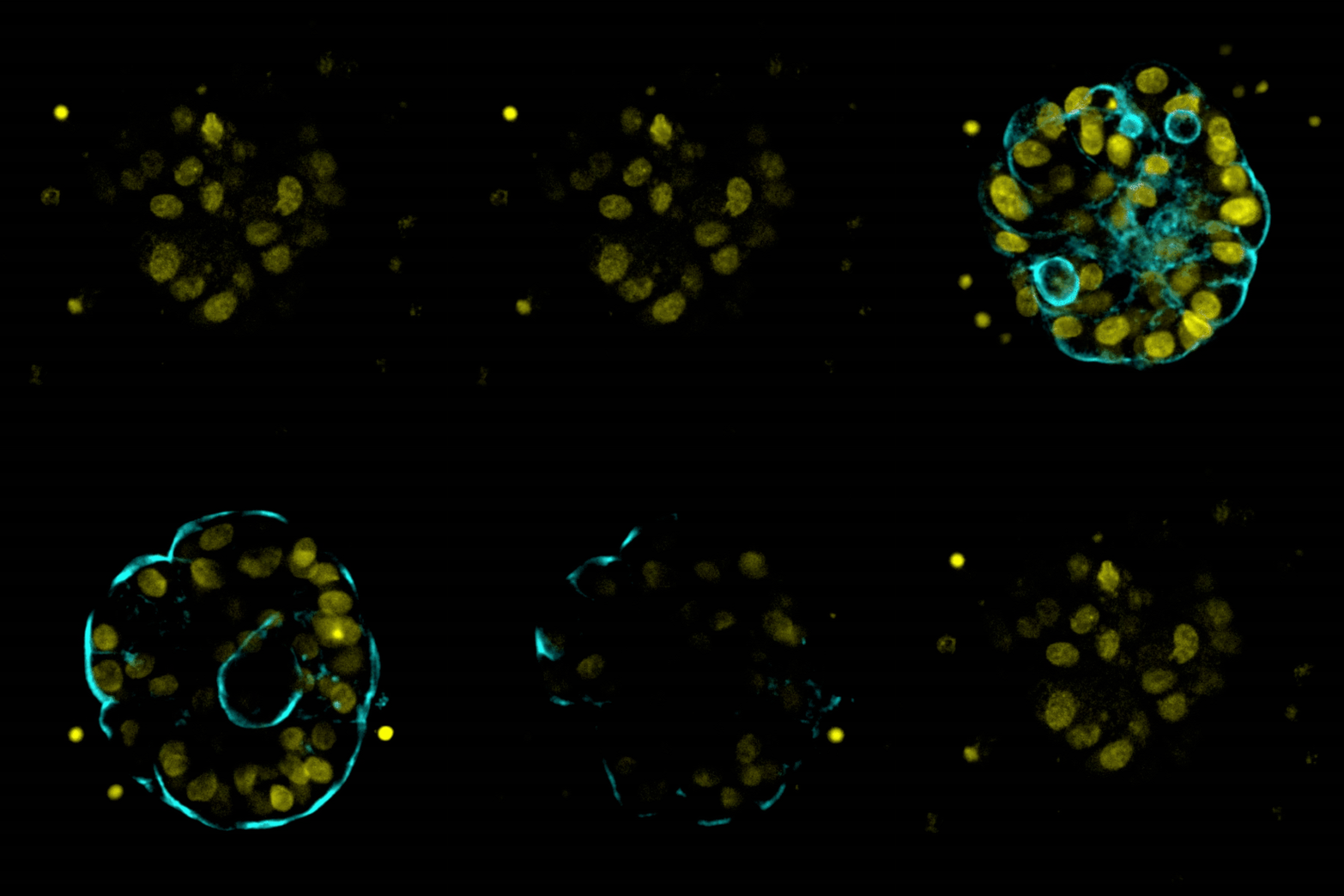
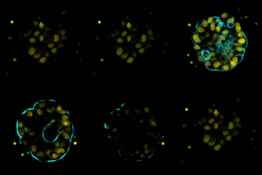
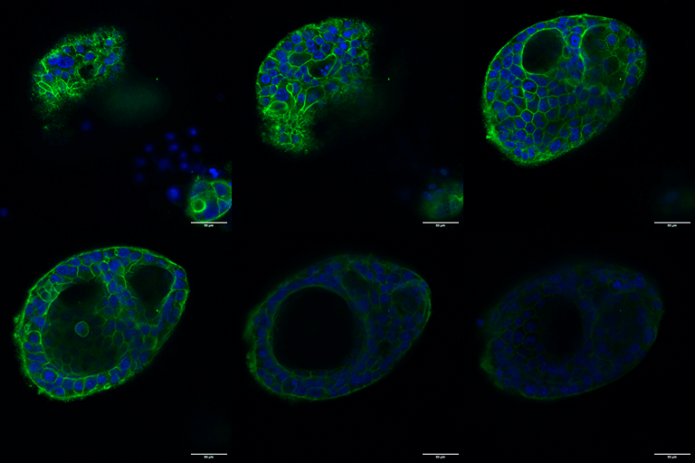
Confocal fluorescence imaging of mouse intestinal organoids.
Mouse intestinal organoids were cultured using RocketCell™ S1 & S2 Supplements, AOF and stained to assess morphology and structural integrity. (A) Nuclear localization visualised by fluorescent staining (yellow). (B) F-actin/membrane visualization highlighting crypt-like budding and luminal architecture (cyan). (C) Merged overlay of nuclear (yellow) and structural (cyan) channels demonstrating organized cellular distribution. (D) Maximum intensity projection / representative z-frame highlighting nuclear density across the 3D organoid volume. Scale bars = 50 µm.
SPheroids
iPSC-derived Neuroectodermal Spheroid Culture in VitroGel® Hydrogels
and the RocketCell™ S1 (B27-like) Supplement, AOF
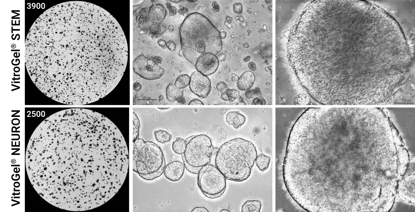
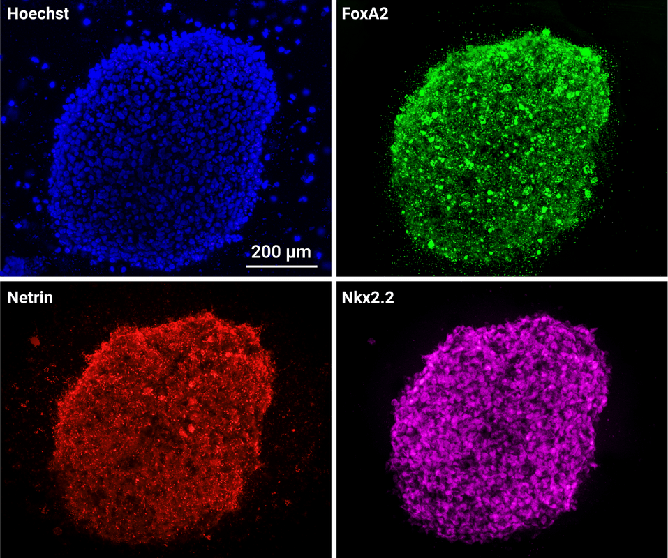
3D iPSC-derived Floor Plate Neuroepithelium Spheroids.
iPSC spheroids cultured with the RocketCell™ 3D iPSC Complete Growth Kit, AOF were transitioned into FPNE1 Medium, and cultured for 4 days, passaged into VitroGel® STEM or VitroGel® NEURON in FPNE medium supplemented with RocketCell™ S1 Supplement, Minus Insulin, AOF and characterized after 8 days. These spheroids expressed canonical FPNE markers such as FoxA2, Netrin, and Nkx2.2.
Floor Plate Neuron Epithelium Spheroid Differentiation into Motor Neurons with the RocketCell™ S1 (B27-like) Supplement, AOF
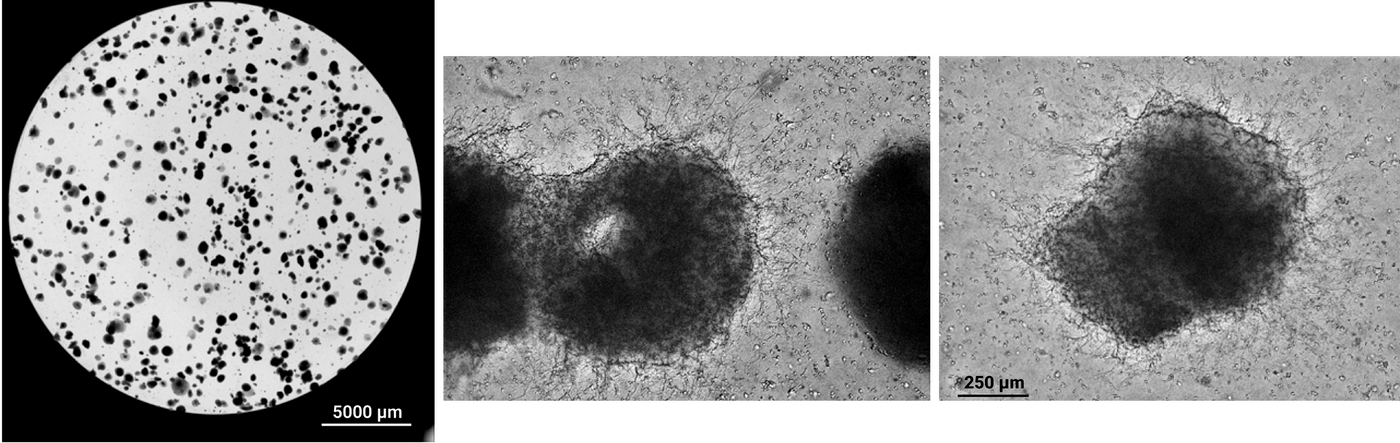
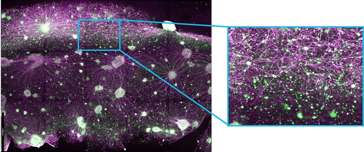
Floor Plate Neuron Epithelium Culture Differentiation into Motor Neurons
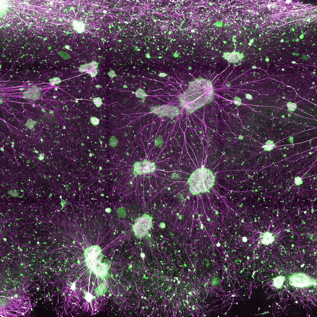
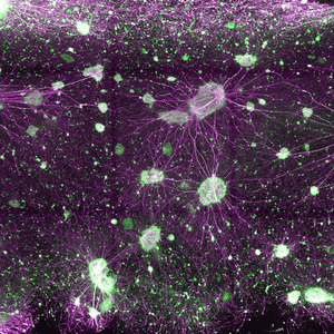
A. FPNE cultures were transitioned to FPNE3 patterning medium for 4 days and transitioned to Neuron Differentiation Medium with the RocketCell™ S1 Supplement, AOF for 14 days.
B. The cultures were fixed and processed for FL immunostaining using anti-phosphorylated neurofilament (purple) and CHAT (green) for motor neurons. This system demonstrates the power of developing complex interconnected systems capable of spanning multi-millimeter volumes in 3D with thousands of interconnected neuron spheroids and interneurons.
| Supplement Type | Standard, Minus Insulin, Minus Vitamin A |
|---|
Related products
New
Cell Culture Media (Animal Origin-Free)
One Core Medium. Endless Organoid Possibilities. An animal origin-free medium that supports multiple organoid types - just add growth factors.
New
Cell Culture Media (Animal Origin-Free)
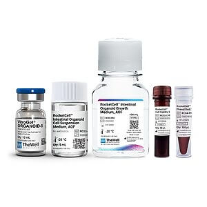
All-in-one, animal origin-free kit for growth, expansion, and passaging of apical-out intestinal organoids.
New

Cell Culture Media (Animal Origin-Free)
$449.00
Animal origin-free growth medium for intestinal organoids
NEW
Culture Media and Supplements
N2-like Animal Origin-Free Supplement
New
Cell Culture Media (Animal Origin-Free)
An all-in-one, animal origin-free kit with optimized matrix, medium, and reagents for 3D iPSC expansion.
New
Cell Culture Media (Animal Origin-Free)
Animal origin-free medium for 2D and 3D expansion of pluripotent stem cells.