
Room Temperature Operation
Room temperature protocol/operation. No ice bucket required.
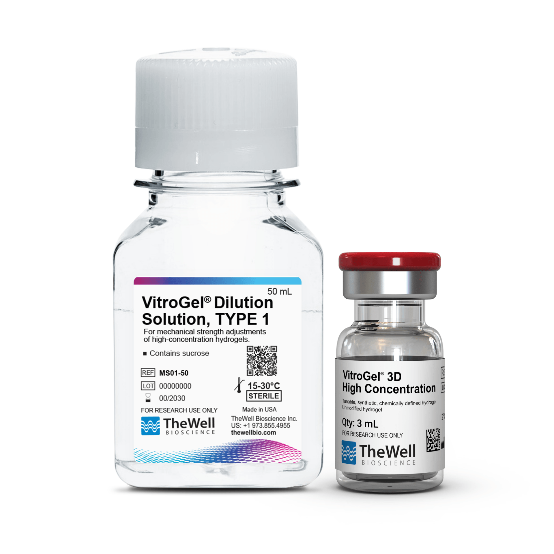
VitroGel® 3D High Concentration
Tunable, synthetic, chemically defined hydrogel, high concentration (3 mL kit)
VitroGel® 3D High Concentration
Tunable, synthetic, chemically defined hydrogel for cell spheroid formation, suspension cells, or cells requiring low cell-matrix interactions.
Synthetic and Chemically Defined
100% synthetic. Animal & human origin-free, biofunctional hydrogel.
Tunable Hydrogel Strength
Adjust the hydrogel strength from 10 Pa to over 20,000 Pa to create the optimal cell environment.

Injectability
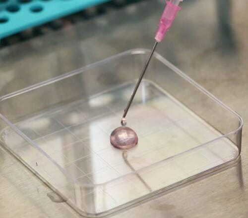
Excellent cell retention. Long-term injectability after gelation without needle clogging.

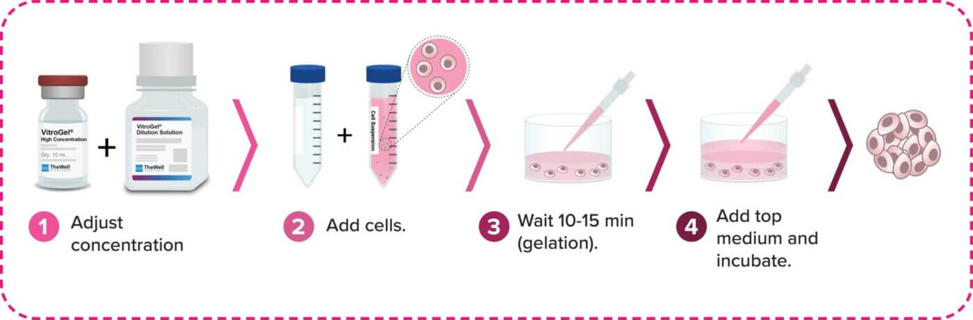
Easy-to-use No cross-linking agent required. Adjust hydrogel with Dilution Solution, mix with cells, add medium and incubate.

Easy cell harvesting Simple and efficient cell harvesting by the non-enzymatic VitroGel® Organoid Recovery Solution.
VitroGel® 3D High Concentration is a tunable, synthetic, and chemically defined hydrogel system that allows the maximum flexibility to manipulate the 3D cell culture environment for different needs. VitroGel® 3D High Concentration comes with VitroGel® Dilution Solution to adjust the final hydrogel strength from 10 to 4000 Pa. The hydrogel’s tunability gives researchers the ability to create an optimized environment for cell growth. The VitroGel® 3D High Concentration hydrogel matrix structure is good for cell spheroid formation, suspension cells, or cells requiring low cell-matrix interactions.
VitroGel® High Concentration hydrogels are our tunable, synthetic and chemically defined hydrogels for researchers wanting full control to manipulate the biophysical and biological properties of the cell culture environment. The tunability of the hydrogel gives the ability to create an optimized environment for cell growth. The hydrogel system has a neutral pH, transparent, permeable, and compatible with different imaging systems. The solution transforms into a hydrogel matrix by simply mixing with the cell culture medium. No cross-linking agent is required. Cells cultured in this system can be easily harvested with our VitroGel® Organoid Recovery Solution. The hydrogel can also be tuned to be injectable for in vivo studies.
From 3D cell culture, 2D cell coating to animal injection, VitroGel® makes it possible to bridge the in vitro and in vivo studies with the same platform system.
3D Cell Culture Process in 20 Minutes
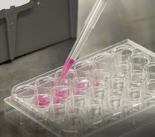
VitroGel® High Concentration hydrogels are easy-to-use. There is no cross-linking agent required. Work confidently at room temperature.
Specifications
| Contents | VitroGel®® 3D High Concentration, 3 mL VitroGel® Dilution Solution, 50 mL |
| Hydrogel Formulation | Synthetic, chemically defined, tunable hydrogel, pure and unmodified. |
| Use | Good for cell spheroid formation, suspension cells or cells require low cell-matrix interactions |
| Mix & Match | Can be blended with other versions of VitroGel® concentrated hydrogels to create a custom multi-functional matrix. |
| Operation | Room temperature |
| Hydrogel Strength | 10 to 4,000 Pa of G’ depending on dilution ratio. Dilute with VitroGel Dilution Solution (TYPE 1 or TYPE 2) for different concentrations. |
| pH | Neutral |
| Color | Transparent |
| Cell Harvesting | VitroGel® Organoid Recovery Solution 5-15 min cell recovery |
| Injectable | Injectable hydrogel |
| Storage | Store at 2-8°C. Ships at ambient temperature |
| Number of Uses | Dilution ratio: 1:2 = 225 uses at 50 µL per well 1:3 = 300 uses at 50 µL per well 1:5 = 450 uses at 50 µL per well |


Data and References
Cell Type Behavior Reference Table – VitroGel® 3D
Studies were performed using VitroGel® 3D in different tissue and cell types.
Cancer/Tumor
| Cell Type | Behavior |
|---|---|
| Brainstem glioma DIPG | Cell proliferation and survival |
| Breast CTC | Cell proliferation |
| Breast T47D | Spheroid formation and proliferation |
| Chordoma Cells | Cell proliferation |
| Hela Cells | Cell proliferation |
| Human osteosarcoma KHOS | Cell proliferation and spheroid formation |
| Human osteosarcoma U2OS | Cell proliferation and spheroid formation |
Epithelial Cells
| Cell Type | Behavior |
|---|---|
| Human NTHY-ORI 3-1 Cells | Enhance spheroids and cluster formation and promote cell viability |
Immune Cells
| Cell Type | Behavior |
|---|---|
| BL5 human beta cells | Enhance spheroids and cluster formation and promote cell viability |
| CD8 + T cells | Enhance spheroids and cluster formation and promote cell viability |
| Priess human lymphoblastoid cells | Enhance spheroids and cluster formation and promote cell viability |
Red Blood Cells
| Cell Type | Behavior |
|---|---|
| Red Blood Cells | Enhance spheroids and cluster formation and promote cell viability |
Stem Cells
| Cell Type | Behavior |
|---|---|
| Human stem cells from apical papilla SCAP | Enhance cell viability |
All Cell Type Behaviors for All VitroGel® Products
| TISSUE/ORGAN TYPE | CELL TYPE | READY TO USE | HIGH CONCENTRATION | BEHAVIOR | |
|---|---|---|---|---|---|
| Beta Cell | BL5 human beta cells | VitroGel® Hydrogel Matrix VitroGel® ORGANOID Disovery Kit | VitroGel® 3D VitroGel® MMP | Enhance spheroids formation | |
| Beta TC3 cells | VitroGel® Hydrogel Matrix VitroGel® ORGANOID Disovery Kit | VitroGel® RGD | Cell proliferation and cellular interactions | ||
| Bone | Bone marrow stromal cells (rat) | VitroGel® Hydrogel Matrix VitroGel® ORGANOID Disovery Kit | VitroGel® RGD VitroGel® COL VitroGel® MMP | Osteogensic differentiation Cell attachment and osteoblast differentiation Cell proliferation cell viability and cellular networking | |
| Osteoblasts (rat) | VitroGel® MSC VitroGel® Hydrogel Matrix VitroGel® ORGANOID Disovery Kit | VitroGel® RGD VitroGel® COL | Cell attachment and spreading | ||
| Bone marrow stromal cells (bovine) | VitroGel® MSC VitroGel® Hydrogel Matrix VitroGel® ORGANOID Disovery Kit | VitroGel® RGD VitroGel® COL | Cell spreading and osteocalcin expression | ||
| Breast | Mammary gland MCF10A | VitroGel® Hydrogel Matrix VitroGel® ORGANOID Disovery Kit | VitrolGel RGD VitroGel® COL VitroGel® MMP | Spheroid formation MMP activity in response to TGF-B1 | |
| Mammary epithelium (mouse) | VitroGel® Hydrogel Matrix VitroGel® ORGANOID Disovery Kit | VitroGel® RGD VitroGel® COL | Cell invasion and dissemination | ||
| Cancer/Tumor | Human colorectal carcinoma HCT 116 | VitroGel® Hydrogel Matrix VitroGel® ORGANOID Disovery Kit | VitroGel® RGD | Cell proliferation cell survival and intercellular networking | |
| Huaman colon carcinoma HCT-8 | VitroGel® Hydrogel Matrix VitroGel® ORGANOID Disovery Kit | VitroGel® RGD | Cell proliferation and cell matrix interaction | ||
| Glioma U87-MG | VitroGel® Hydrogel Matrix VitroGel® ORGANOID Disovery Kit | VitroGel® RGD VitroGel® MMP VitroGel® COL | Cell spreading and acting stress fiber assembly cell proliferation spreading and migration Cell migration dependent on mechancial force Cell proliferation and cell matrix interaction | ||
| Gliobastoma SF 268 | VitroGel® Hydrogel Matrix VitroGel® ORGANOID Disovery Kit | VitrolGel RGD | Cell proliferation and cell matrix interaction | ||
| Gliobastoma SF 295 | VitroGel® Hydrogel Matrix VitroGel® ORGANOID Disovery Kit | VitroGel® RGD | Cell proliferation and cell matrix interaction | ||
| Glioblastoma SNB75 | VitroGel® Hydrogel Matrix VitroGel® ORGANOID Disovery Kit | VitroGel® RGD | Cell proliferation and cell matrix interaction | ||
| Glioblastoma U-251 MG | VitroGel® Hydrogel Matrix VitroGel® ORGANOID Disovery Kit | VitroGel® RGD | Cell proliferation and cell matrix interaction | ||
| Prostate PC3 | VitroGel® Hydrogel Matrix VitroGel® ORGANOID Disovery Kit | VitroGel® COL VitroGel® IKVAV VitroGel® RGD VitroGel® MMP | Cell proliferation reduced MMP release invasion migration and spheroid metabolic activity. | ||
| Prostate LNCaP | VitroGel® Hydrogel Matrix VitroGel® ORGANOID Disovery Kit | VitroGel® RGD VitroGel® COL | Cell attachment proliferation and prostate specific antigen release | ||
| Prostate CRPC | VitroGel® Hydrogel Matrix VitroGel® ORGANOID Disovery Kit | VitroGel® RGD | Cell proliferation and invasion | ||
| Prostate DU145 | VitroGel® Hydrogel Matrix VitroGel® ORGANOID Disovery Kit | VitroGel® RGD | Cell proliferation and invasion | ||
| Melanoma B16F10 | VitroGel® Hydrogel Matrix VitroGel® ORGANOID Disovery Kit | VitroGel® COL VitroGel® YIGSR | Cell migration invasion MMP release cell attachment and spreading | ||
| Breast MDA-MB-231 | VitroGel® Hydrogel Matrix VitroGel® ORGANOID Disovery Kit | VitroGel® RGD VitroGel® MMP VitroGel® 3D | Cell invasion spreading proliferation division migration and cluster growth | ||
| Fibrosarcoma HT1080 | VitroGel® Hydrogel Matrix VitroGel® ORGANOID Disovery Kit | VitroGel® RGD VitroGel® COL | Cell infiltration attachment | ||
| Breast T47D | VitroGel® Hydrogel Matrix VitroGel® ORGANOID Disovery Kit | VitroGel® COL VitroGel® 3D VitroGel® RGD VitroGel® MMP | Force dependent tubule formation cell cluster growth spheroid formation and proliferation | ||
| Breast 4T1 | VitroGel® Hydrogel Matrix VitroGel® ORGANOID Disovery Kit | VitroGel® RGD | Cell proliferation | ||
| Breast CTC | VitroGel® Hydrogel Matrix VitroGel® ORGANOID Disovery Kit | VitroGel® 3D VitroGel® RGD | Cell proliferation | ||
| Breast E0771 | VitroGel® Hydrogel Matrix VitroGel® ORGANOID Disovery Kit | VitroGel® RGD | Cell proliferation spheroid formation | ||
| Brest AU-565 | VitroGel® Hydrogel Matrix VitroGel® ORGANOID Disovery Kit | VitroGel® RGD | Cell proliferation cell matrix interactions | ||
| Epithelial ovarian OV-MZ-6 | VitroGel® Hydrogel Matrix VitroGel® ORGANOID Disovery Kit | VitroGel® RGD | Spheroid formation and proliferation | ||
| Epithelial ovarian SKOV-3 | VitroGel® Hydrogel Matrix VitroGel® ORGANOID Disovery Kit | VitroGel® RGD | Spheroid formation and proliferation | ||
| Glioma U373-MG | VitroGel® Hydrogel Matrix VitroGel® ORGANOID Disovery Kit | VitroGel® RGD VitroGel® COL VitroGel® MMP | Cell adhesion invasion and migration | ||
| Rhabdomyosarcoma (human) | VitroGel® Hydrogel Matrix VitroGel® ORGANOID Disovery Kit | VitroGel® RGD VitroGel® COL VitroGel® YIGSR | Cell attachment and spreading | ||
| Melanoma SK-MEL-28 | VitroGel® Hydrogel Matrix VitroGel® ORGANOID Disovery Kit | VitroGel® RGD VitroGel® COL VitroGel® IKVAV | Cell adhesion and proliferation | ||
| Melanoma K-1735 | VitroGel® Hydrogel Matrix VitroGel® ORGANOID Disovery Kit | VitroGel® RGD VitroGel® COL VitroGel® IKVAV | Cell invasion | ||
| Melanoma A2058 | VitroGel® Hydrogel Matrix VitroGel® ORGANOID Disovery Kit | VitroGel® RGD VitroGel® COL VitroGel® IKVAV | Collagenolytic activity | ||
| Brainstem glioma DIPG | VitroGel® Hydrogel Matrix VitroGel® ORGANOID Disovery Kit | VitroGel® RGD VitroGel® COL VitroGel® MMP | Cell proliferation and survival | ||
| Hela Cells | VitroGel® Hydrogel Matrix VitroGel® ORGANOID Disovery Kit | VitroGel® 3D VitroGel® RGD VitroGel® MMP | Cell proliferation | ||
| Colorectal adenocarcinoma DLD-1 cells | VitroGel® Hydrogel Matrix VitroGel® ORGANOID Disovery Kit | VitroGel® RGD | Cell proliferation and cell matrix interaction | ||
| Giloma LRM55 | VitroGel® Hydrogel Matrix VitroGel® ORGANOID Disovery Kit | VitroGel® RGD VitroGel® IKVAV VitroGel® MMP | Cell attachment | ||
| Melanoma WM 239A | VitroGel® Hydrogel Matrix VitroGel® ORGANOID Disovery Kit | VitroGel® RGD VitroGel® COL VitroGel® MMP | Cell invasion | ||
| Melanoma Cells | VitroGel® Hydrogel Matrix VitroGel® ORGANOID Disovery Kit | VitroGel® RGD | Cell proliferation and cell matrix interaction | ||
| Insulinoma ins-1 (Rat) | VitroGel® Hydrogel Matrix VitroGel® ORGANOID Disovery Kit | VitroGel® RGD | Cell proliferation and cell matrix interaction | ||
| Biphasic synovial sarcoma SYO-1 | VitroGel® Hydrogel Matrix VitroGel® ORGANOID Disovery Kit | VitroGel® RGD | Cell proliferation cell matrix interation and cell survival | ||
| Fuji Cells | VitroGel® Hydrogel Matrix VitroGel® ORGANOID Disovery Kit | VitroGel® RGD | Cell proliferation and cell matrix interaction | ||
| Chordoma Cells | VitroGel® Hydrogel Matrix VitroGel® ORGANOID Disovery Kit | VitroGel® 3D | Cell proliferation | ||
| Bone OSA 1777 | VitroGel® Hydrogel Matrix VitroGel® ORGANOID Disovery Kit | VitroGel® RGD | Spheroid and cluster formation | ||
| Glioma RuGli | VitroGel® Hydrogel Matrix VitroGel® ORGANOID Disovery Kit | VitroGel® COL | Integrin dependent cell adhesion | ||
| Breast Cancer MCF-7 | VitroGel® Hydrogel Matrix VitroGel® ORGANOID Disovery Kit | VitroGel® RGD VitroGel® COL VitroGel® MMP VitroGel® 3D | Cell proliferation intercellular connections morphological changes MMP expression and angiogenesis | ||
| Liver carcinoma HepG2 | VitroGel® Hydrogel Matrix VitroGel® ORGANOID Disovery Kit | VitroGel® RGD VitroGel® COL | Cell viability growth drug resistance proliferation and cellular matrix interaction | ||
| Human pancreatic cancer PANC-1 | VitroGel® Hydrogel Matrix VitroGel® ORGANOID Disovery Kit | VitroGel® RGD VitroGel® COL VitroGel® MMP | Cell proliferation and cellular interactions | ||
| Primary breast (human) | VitroGel® Hydrogel Matrix VitroGel® ORGANOID Disovery Kit | VitroGel® RGD VitroGel® COL VitroGel® MMP | Cell invasion migration and dissemination | ||
| Ovarian carcinoma OVCAR-3 | VitroGel® Hydrogel Matrix VitroGel® ORGANOID Disovery Kit | VitroGel® RGD VitroGel® MMP | Cell proliferation cell matrix interactions | ||
| Ovarian OVCA429 | VitroGel® Hydrogel Matrix VitroGel® ORGANOID Disovery Kit | VitroGel® RGD VitroGel® MMP VitroGel® COL | MMP dependent cell invasion | ||
| Human Osteosarcoma KHOS | VitroGel® Hydrogel Matrix VitroGel® ORGANOID Disovery Kit | VitroGel® RGD VitroGel® 3D | Cell proliferation and spheroids formation | ||
| Human Osteosarcoma U2OS | VitroGel® Hydrogel Matrix VitroGel® ORGANOID Disovery Kit | VitroGel® RGD VitroGel® 3D | Cell proliferation and spheroids formation | ||
| Human fibroblast-like synoviocytes (FLS) | VitroGel® Hydrogel Matrix VitroGel® ORGANOID Disovery Kit | VitroGel® 3D | Cell proliferation and inflammatory responses | ||
| Human Liposarcoma 94T778 | VitroGel® Hydrogel Matrix VitroGel® ORGANOID Disovery Kit | VitroGel® 3D | Cell proliferation and spheroids formation | ||
| Human diffuse large B-cell lymphoma (DLBLC) SUDHL-10 | VitroGel® Hydrogel Matrix | Cell viability growth drug resistance proliferation and cellular matrix interaction | |||
| Priess human lymphoblastoid cells | VitroGel® Hydrogel Matrix VitroGel® ORGANOID Disovery Kit | VitroGel® 3D | Enhance spheroids and cluster formation and promote cell viability. | ||
| Cartilage | Chondrocytes (bovine) | VitroGel® MSC VitroGel® Hydrogel Matrix VitroGel® ORGANOID Disovery Kit | VitroGel® RGD VitroGel® COL | Cell viability and proliferation | |
| Chondrocytes (human) | VitroGel® MSC VitroGel® Hydrogel Matrix VitroGel® ORGANOID Disovery Kit | VitroGel® RGD VitroGel® COL | Cell viability and proliferation | ||
| Connective Tissue | Dermal Fibroblasts (human) | VitroGel® Hydrogel Matrix VitroGel® ORGANOID Disovery Kit | VitroGel® RGD | Cell viability and spreading | |
| Fibroblasts NIH3T3 | VitroGel® Hydrogel Matrix VitroGel® ORGANOID Disovery Kit | VitroGel® RGD VitroGel® COL | Directional cell migration toward gradient and cell spreading dependent on substrata rigidity | ||
| Foreskin fibroblasts (human) | VitroGel® Hydrogel Matrix VitroGel® ORGANOID Disovery Kit | VitroGel® RGD VitroGel® COL VitroGel® YIGSR VitroGel® MMP | Cell spreading substrata degradation and cell invasion | ||
| Skin fibroblasts (skin) | VitroGel® Hydrogel Matrix VitroGel® ORGANOID Disovery Kit | VitroGel® RGD VitroGel® IKVAV | Cell adhesion | ||
| Epidermal keratinocytes | VitroGel® Hydrogel Matrix VitroGel® ORGANOID Disovery Kit | VitroGel® RGD VitroGel® COL | Cell viability | ||
| Epithelial Cells | Mouse ovarian follicle cells | VitroGel® Hydrogel Matrix VitroGel® ORGANOID Disovery Kit | VitroGel® RGD | 3D cell culture using ES-hydrogel can enhance vitro follicle culture by considering the permeability and stiffness of the gel. | |
| Human Nthy-ori 3-1 cells | VitroGel® Hydrogel Matrix VitroGel® ORGANOID Disovery Kit | VitroGel® 3D | Enhance spheroids and cluster formation and promote cell viability. | ||
| A549 cells | VitroGel® Hydrogel Matrix VitroGel® ORGANOID Disovery Kit | VitroGel® RGD | Enhance cell proliferation and cell matrix interactions. | ||
| MCF-12A | VitroGel® Hydrogel Matrix VitroGel® ORGANOID Disovery Kit | VitroGel® RGD | Enhance cell proliferation and cell matrix interactions. | ||
| Immortalized bronchial epithelial cells HBEC-KRAS | VitroGel® Hydrogel Matrix VitroGel® ORGANOID Disovery Kit | VitroGel® RGD VitroGel® 3D | Cell proliferation | ||
| Eye | Corneal endothelial B4G12 | VitroGel® Hydrogel Matrix VitroGel® ORGANOID Disovery Kit VitroGel® Angiogenesis Assay | VitroGel® RGD VitroGel® Angiogenesis Assay HC kit | Cell attachment and spreading | |
| Retinal ganglion cells (xenopus) | VitroGel® Hydrogel Matrix VitroGel® ORGANOID Disovery Kit | VitroGel® RGD VitroGel® COL | Neurite outgrowth | ||
| Immune Cells | CD8 + T cells | VitroGel® Hydrogel Matrix VitroGel® ORGANOID Disovery Kit | VitroGel® 3D | Enhance spheroids and cluster formation and promote cell viability. | |
| Kidney | Human embryonic kidney HEK293 | VitroGel® Hydrogel Matrix VitroGel® HEK293 VitroGel® ORGANOID Disovery Kit | VitroGel® RGD VitroGel® COL | 3D spheroids formation | |
| Madin-Darby Canine Kidney | VitroGel® Hydrogel Matrix VitroGel® HEK293 VitroGel® ORGANOID Disovery Kit | VitroGel® RGD VitroGel® MMP | Epithelial cysts formation | ||
| Podocytes (human) | VitroGel® Hydrogel Matrix VitroGel® HEK293 VitroGel® ORGANOID Disovery Kit | VitroGel® RGD VitroGel® COL | Glomerular capillary formation | ||
| glomerular endothelial cells (human) | VitroGel® Hydrogel Matrix VitroGel® HEK293 VitroGel® ORGANOID Disovery Kit VitroGel® Angiogenesis Assay | VitroGel® RGD VitroGel® Angiogenesis Assay HC kit | Glomerular capillary formation | ||
| Liver | Hepatocytes (human) | VitroGel® Hydrogel Matrix VitroGel® ORGANOID Disovery Kit | VitroGel® RGD VitroGel® COL | Filopodia formation and synthesis of albumin and cell attachment | |
| Hepatocytes (mouse rat swine) | VitroGel® Hydrogel Matrix VitroGel® ORGANOID Disovery Kit | VitroGel® RGD VitroGel® COL VitroGel® MMP | Cell viability spearding Albumin secretion | ||
| Lung | Alveolar basal epithelial A549 | VitroGel® Hydrogel Matrix VitroGel® ORGANOID Disovery Kit | VitroGel® RGD | Cell attachment | |
| Alveolar epithelial RLE-6TN | VitroGel® Hydrogel Matrix VitroGel® ORGANOID Disovery Kit | VitroGel® RGD | Cell attachment and mesenchymal differentiation | ||
| Pulmonary fibroblasts LL2 | VitroGel® Hydrogel Matrix VitroGel® ORGANOID Disovery Kit | VitroGel® RGD VitroGel® IKVAV | Cell adhesion | ||
| HFL1 lung fibroblasts CCL153 | VitroGel® Hydrogel Matrix VitroGel® ORGANOID Disovery Kit | VitroGel® RGD | Cell proliferation and spindle morphology | ||
| Lung cancer associated fibroblasts (human) | VitroGel® Hydrogel Matrix VitroGel® ORGANOID Disovery Kit | VitroGel® RGD | Substrata contractility | ||
| Lung fibroblasts MCR-5 | VitroGel® Hydrogel Matrix VitroGel® ORGANOID Disovery Kit | VitroGel® RGD VitroGel® COL | NGF-mediated substrata contraction | ||
| Muscle | Myoblasts C2C12 | VitroGel® Hydrogel Matrix VitroGel® ORGANOID Disovery Kit | VitroGel® RGD VitroGel® COL | Cell proliferation differentiation attachment myofibril formation myotube formation and integrin dependent cell adhesion | |
| Skeletal myoblasts (mouse) | VitroGel® Hydrogel Matrix VitroGel® ORGANOID Disovery Kit | VitroGel® RGD | Cell attachment proliferation and myofibril formation | ||
| Myoblasts (human) | VitroGel® Hydrogel Matrix VitroGel® ORGANOID Disovery Kit | VitroGel® RGD VitroGel® COL | Cell adhesion alignment along fiber and myotube formation | ||
| Myoblasts C25Cl48 | VitroGel® Hydrogel Matrix VitroGel® ORGANOID Disovery Kit | VitroGel® RGD VitroGel® COL | Cell proliferation differentiation and myotube formation | ||
| Neural | Dorsal root ganglion (chick) | VitroGel® Hydrogel Matrix VitroGel® ORGANOID Disovery Kit VitroGel® NEURON | VitroGel® RGD VitroGel® COL | Neurite formation and force dependent neurite outgrowth | |
| Neural PC12 | VitroGel® Hydrogel Matrix | VitroGel® RGD VitroGel® IKVAV | Neurite outgrowth | ||
| Neural stem cell/ progenitor cell (rat) | VitroGel® STEM VitroGel® Hydrogel Matrix VitroGel® NEURON | VitroGel® RGD VitroGel® IKVAV | Cell viability attachment and differentiation | ||
| Neural stem cell/ progenitor cell (human) | VitroGel® STEM VitroGel® Hydrogel Matrix VitroGel® NEURON | VitroGel® RGD VitroGel® IKVAV VitroGel® COL | Cell viability attachment and differentiation | ||
| Schwann cells (rat) | VitroGel® Hydrogel Matrix VitroGel® NEURON | VitroGel® RGD | Cell attachment and migration | ||
| Neural stem cell/ progenitor cell (mouse) | VitroGel® STEM VitroGel® NEURON | VitroGel® RGD VitroGel® IKVAV | Cell adhesion and differentiation | ||
| Cortical astrocytes (rat) | VitroGel® Hydrogel Matrix VitroGel® NEURON | VitroGel® RGD VitroGel® IKVAV | Cell adhesion | ||
| Spiral ganglion neurons (mouse) | VitroGel® Hydrogel Matrix VitroGel® NEURON | VitroGel® RGD VitroGel® IKVAV | Neurite outgrowth | ||
| Motor neurons (human) | VitroGel® Hydrogel Matrix VitroGel® NEURON | VitroGel® RGD VitroGel® COL VitroGel® IKVAV | Force dependent neurite outgrowth | ||
| Forebrain neurons (human) | VitroGel® Hydrogel Matrix VitroGel® NEURON | VitroGel® RGD VitroGel® COL VitroGel® IKVAV | Force dependent neurite outgrowth | ||
| Cortical neurons (rat) | VitroGel® Hydrogel Matrix VitroGel® NEURON | VitroGel® RGD VitroGel® COL VitroGel® IKVAV | Neuronal viability and neurite outgrowth | ||
| Dorsal root ganglion (rat) | VitroGel® Hydrogel Matrix VitroGel® NEURON | VitroGel® RGD VitroGel® COL VitroGel® IKVAV | Neurite outgrowth | ||
| Red Blood Cells | Red Blood Cells | VitroGel® Hydrogel Matrix VitroGel® ORGANOID Disovery Kit | VitroGel® 3D | Enhance Spheroids and cluster formation and promote cell viability | |
| Pancreas | B-cells MIN6 | VitroGel® Hydrogel Matrix VitroGel® ORGANOID Disovery Kit | VitroGel® RGD VitroGel® IKVAV | Reduced apoptosis and increased insulin release | |
| Stem Cells | Mesenchymal stem cells (human) | VitroGel® MSC | VitroGel® RGD VitroGel® COL VitroGel® IKVAV VitroGel® MMP | Cell viability proliferation differentiation neuronal differntiation neurite outgrowth attachment spreading viability and osteoblast differentiation | |
| Mesenchymal stem cells (mouse) | VitroGel® MSC | VitroGel® RGD VitroGel® MMP | Cell spreading and migration | ||
| Mesenchymal stem cells (rat) | VitroGel® MSC | VitroGel® RGD | Cell adhesion and spreading | ||
| Embryonic stem cells (mouse) | VitroGel® STEM VitroGel® ORGANOID Disovery Kit | VitroGel® RGD VitroGel® COL VitroGel® YIGSR | Endothelial cell differentiation neuronal differentiation and neurite outgrowth | ||
| Induced pluripotent stem cells (human) | VitroGel® STEM VitroGel® ORGANOID Disovery Kit | VitroGel® RGD VitroGel® YIGSR VitroGel® IKVAV | Cell viability | ||
| Human stem cells from apical papilla SCAP | VitroGel® STEM | VitroGel® RGD | Cell viability | ||
| Hematopoietic Stem Cells | VitroGel® STEM | Cell viability | |||
| Adipose derived stem cells (human) | VitroGel® MSC | VitroGel® RGD VitroGel® 3D VitroGel® IKVAV | Cell viability cell attachment | ||
| Vascular/cardiac | Umbilical vein endothelial cells | VitroGel® Angiogenesis Assay VitroGel® Hydrogel Matrix VitroGel® ORGANOID Disovery Kit | VitroGel® Angiogenesis Assay HC kit | Cell attachment proliferation migration angiogenesis gene expression changes migratory cell infiltration cell survival and VEGF dependent migration | |
| Neonatal cardiac (rat) | VitroGel® Angiogenesis Assay VitroGel® Hydrogel Matrix VitroGel® ORGANOID Disovery Kit | VitroGel® Angiogenesis Assay HC kit | Cell attachment tissue regeneration and attachment similar to laminin | ||
| Aortic smooth muscle cells | VitroGel® Hydrogel Matrix | VitroGel® Angiogenesis Assay HC kit | Cell attachment | ||
| Endothelial (human) | VitroGel® Angiogenesis Assay | VitroGel® Angiogenesis Assay HC kit | Cell differentiation | ||
| Endotheliocytes | VitroGel® Angiogenesis Assay | VitroGel® Angiogenesis Assay HC kit | Cell migration | ||
| Microvascular endothelial cells (human) | VitroGel® Angiogenesis Assay | VitroGel® Angiogenesis Assay HC kit | Cell mobility | ||
| Aortic endothelial cells (bovine) | VitroGel® Angiogenesis Assay | VitroGel® Angiogenesis Assay HC kit | Force dependent cell spreading | ||
| Capillary endothelial cells (bovine) | VitroGel® Angiogenesis Assay | VitroGel® Angiogenesis Assay HC kit | Capillary like network formation |
Data
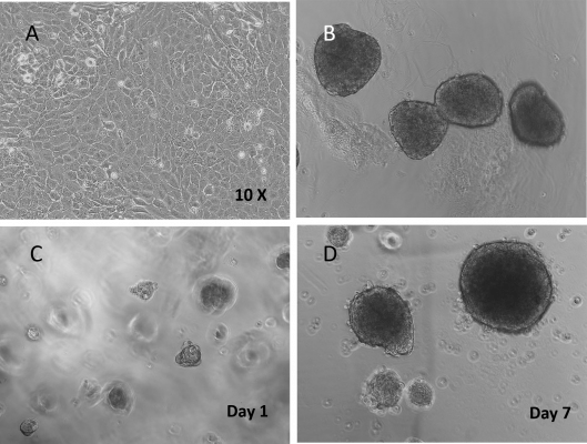
Figure 1. Beta Lox 5 (BL5) cells 3D culture in VitroGel® 3D system.
A. BL5 cells cultured on the surface of a regular tissue culture-treated well plate (control); B. Normal human islets grown in suspension culture (comparison); C. 3D culture of BL5 cells in VitroGel 3D at Day 1; D. 3D culture of BL5 cells in VitroGel 3D at Day 7. Under 3D culture of VitroGel 3D, BL5 cells form islet-like structures very similar to normal human islets. The hydrogel is prepared at 1:3 dilution. The images were taken at 10X magnification.
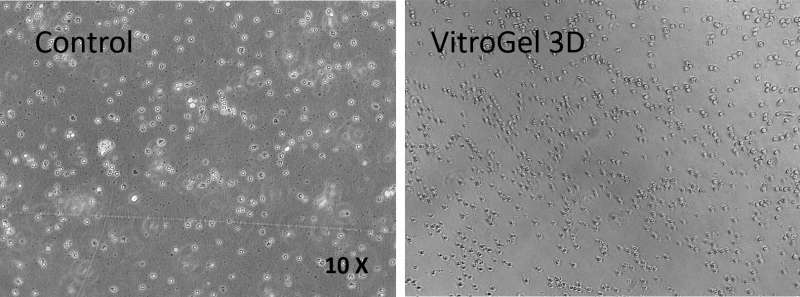
Figure 2. CD8+ T cells 3D culture in VitroGel® 3D system.
CD8+ T cells culture grew in suspension culture (control); B. 3D culture of CD8+ T cells in VitroGel 3D at Day 7. CD8+ T cells are vibrant in 3D culture conditions of VitroGel 3D. The cells can easily move within the unmodified hydrogel matrix. The hydrogel is prepared at 1:3 dilution. The images were taken at 10X magnification.
2D Coating Applications
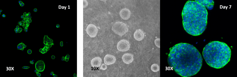
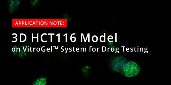
Figure 3. Human colon cancer cells (HCT 116) cells cultured on top of VitroGel® 3D hydrogel.
A thick hydrogel coating plate has been prepared by mixing VitroGel® 3D with PBS at 1:1 ratio. A 300 µL mixture has been added to a well of a 24-well plate and stabilization at room temperature for 20 minutes before adding cells on top of the hydrogel. Cell spheroids form on top of the hydrogel. Cells seeded at 2.5-10×105 cells/mL.
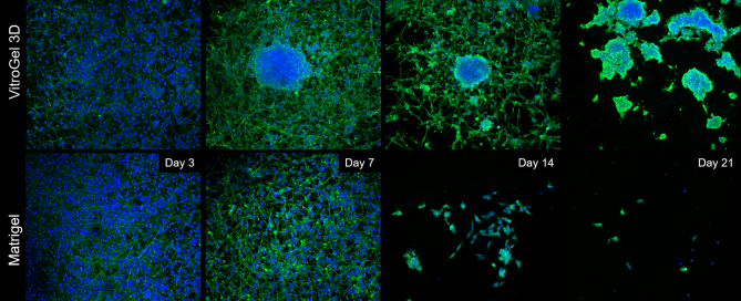
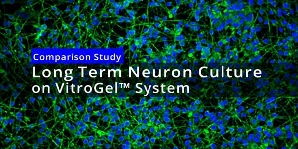
Figure 4. Comparison of long-term neuronal culture seeded onto thick hydrogel mats.
Cells are stained with Beta-III-Tubulin (green) cytoskeleton marker and their nuclei are counter-stained with DAPI (blue). Cells spread out and form neural-like networks as early as day 3 post-differentiation, with comparable efficacy between VitroGel 3D and Matrigel, based on cell survival, culture spreading, and morphological analysis, reached between days 7 and 9. On Matrigel mats, cell culture health and viability drop off sharply once day 9 has passed, with most cells detaching and neurites retracting by day 14, and the vast majority of cells gone by day 21. If grown onto VitroGel® 3D mats, differentiated B35 neurons have a tendency to self-organize into 3D clusters very early on (Day 7), assuming a mixed 2D/3D cell culture for the first two weeks of the time course. By Day 21, these cells have migrated into self-assembled 3D clusters, embedded into the thick hydrogel matrix, with very few cells between the clusters, but without any significant cell death.
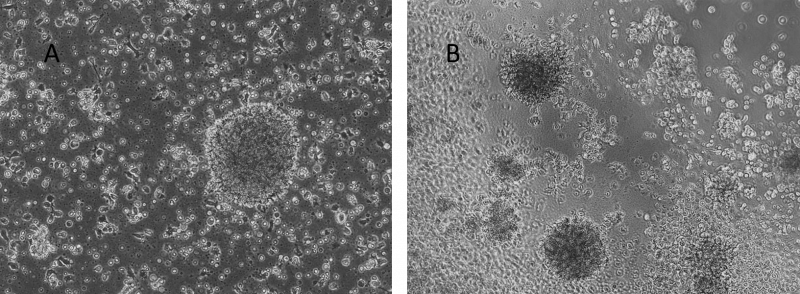
Figure 5. Human Lymphoblastoid Priess cells cultured on top of VitroGel® 3D hydrogel.
A. Priess cells grown in suspension (control); B. Priess cells grown on top of VitroGel® 3D at day 7. A hydrogel substance can be prepared with different stiffness by adjusting the dilution of VitroGel 3D from 1:1 to 1:3 ratio. Cells seeded on the top of the hydrogel form cell spheroids form on the top of the hydrogel. The hydrogel provides a soft substance for cells to attach and grow.
Video Protocols & Demonstrations
Application Notes
Application Notes
Long-Term Neuron Culture Maturation in 3D Hydrogel Constructs
Application Notes
3D Cell Culture of Human Colon Cancer Cells (HCT116) on VitroGel® System
Application Notes
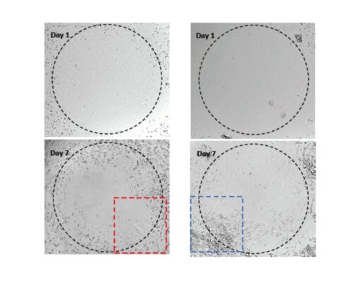
3D Invasion of Glioblastoma Cells in VitroGel® Hydrogel System
Research Highlights
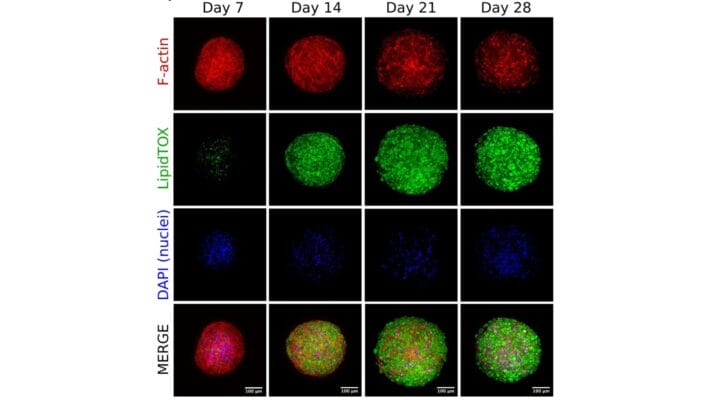
Research Highlights
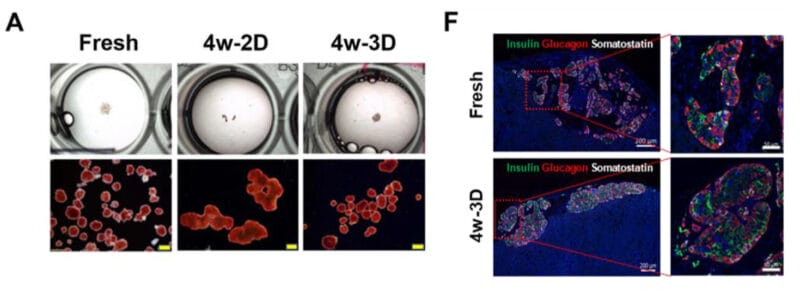
Long-Lived Lab Grown Cells for Diabetes Treatment
Research Highlights
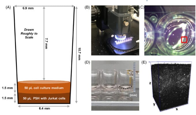
Finding Living Needles in a 3D Haystack
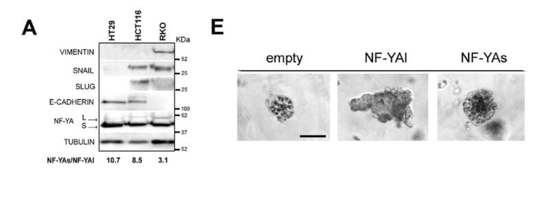
Research Highlights
Cutting Colon Cancer Out Properly

Research Highlights
Go With the PVP Flow for Invitro Model of Hydrocephalus
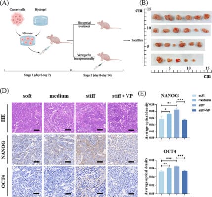
Research Highlights
No Relaxing in the Fight Against Liver Cancer
References/Publications
- Sun, W., Yang, B., Zheng, H., Wang, Q., Shi, Y., Wang, G., Yin, W., Wang, H., Qi, X., Yao, Y., Wang, J., Cui, S., Cui, K., Mao, J., Xia, Y., Deng, J., Zhao, S., Wang, Z., Liu, F., … Zheng, J. (2026). Extracellular matrix remodeling upregulates hippocampal neurogenic niche stiffness and impairs neurogenesis in alzheimer’s disease. Alzheimer’s & Dementia, 22(9). https://doi.org/10.1002/alz.71784
- Mohsen Farrokhpour, Safa Samadzadeh Etehadi, Pejman Hassanpoor, Faraj, T. A., Hasan, A. H., Vesal Abbasian, Sargol Aminnezhad, Azimi, M., Kashanian, S., & Alavi, M. (2026). The promising applications of 3D printing technology for diagnosis and therapy of cancer: Recent advances and challenges. BioImpacts, 16(1), 32895–32895. https://doi.org/10.34172/bi.32895
- Beco, M., Di Pasquale, F., Valenti, C., Betti, P., Mascolo, G. L., Marinucci, L., Eramo, S., & Pagano, S. (2026). Pulp–Dentin Regeneration via Cell Homing: Current Evidence and Perspectives on Cell-Free Regenerative Endodontic Therapy. Medicina, 62(2), 375. https://doi.org/10.3390/medicina62020375
- Andrade, D. C., Salazar-Ardiles, C., Toledo, C., Bueno, J., Cabrera, A. P., Diaz-Jara, E., María Rodriguez-Fernandez, Millet, G. P., Iturriaga, R., & Kelley, E. F. (2025). Chemogenetic inhibition of the carotid bodies blunts hind-limb suspension microgravity-induced muscle alterations in rats. American Journal of Physiology-Lung Cellular and Molecular Physiology. https://doi.org/10.1152/ajplung.00386.2025
- Acimovic, I., Chochola, V., Herrera, J. L., Hampl, A., & Jaros, J. (2025). 3D endothelial network formation in hydrogels improved by stromal cells and specific growth factors. Scientific Reports, 15(1). https://doi.org/10.1038/s41598-025-25381-x
- Meher Beigi Masihi, Chambers, K. R., Friedman, R., Sergey Pampou, Gudenas, B. L., Torrejon, J., Lee, C., Furnari, G., Chau, L. Q., Sajina GC, Lin, Y., Chapman, O. S., Skowron, P., Garzia, L., Taylor, M. D., Chavez, L., Olivier Ayrault, Northcott, P. A., Karan, C., & Wechsler-Reya, R. J. (2025). Ras-Responsive Element Binding Protein 1 regulates survival of Group 3 medulloblastoma. BioRxiv (Cold Spring Harbor Laboratory). https://doi.org/10.1101/2025.07.16.665230
- Yan, Q., Xu, C., Gong, L., Liang, D., Yang, J., Zheng, Y., & Wang, J. (2025). The role of ZC3H13 in promoting M2 macrophage infiltration via m6A methylation in esophageal squamous cell carcinoma tumor progression. Frontiers in Immunology, 16. https://doi.org/10.3389/fimmu.2025.1612041
- Silva, Ferreira, M. C., Grazziotin-Soares, R., Dourado, L. G., Claudia, Rodrigues, G., & Carvalho, C. N. (2025). Regenerative endodontic procedure using Emdogain: a case series. Journal of Medical Case Reports, 19(1). https://doi.org/10.1186/s13256-025-05199-x
- Andrade, D. C., Salazar‐Ardiles, C., Toledo, C., Alvarez, C., Díaz‐Jara, E., Rodriguez‐Fernandez, M., Millet, G. P., & Iturriaga, R. (2025). The carotid body mediates peak oxygen uptake during maximal physical exertion in rats. The Journal of Physiology. https://doi.org/10.1113/jp288633
- Chen, A. R., Chansky, J., & Burke, J. A. (2025). Long-term storage, cryopreservation, and culture of isolated human islets: a systematic review. Frontiers in Transplantation, 4. https://doi.org/10.3389/frtra.2025.1614849
- Zhou, H., Zhang, Z., Liu, Z., Sa, G., Jiang, M., Zou, Z., Shi, Y., Zheng, L., Yang, X., & Sa, G. (2025). Single‐Cell Analysis Reveals Fibroblast‐Derived Migrasomes as CXCL12 Carriers Promoting Skin Wound Repair. Journal of Extracellular Vesicles, 14(6). https://doi.org/10.1002/jev2.70112
- Sun, P., Qin, W., Xu, H., Yin, H., Yang, L., Zhang, X., Jin, X., Xu, Q., Wu, H., Xiaoling Kuai, Jia, L., Huang, J., & Wang, Y. (2025). SPTSSA facilitates gastric cancer progression with modulating PD-L1 in immunomicroenvironment through Wnt/β-catenin pathway. Cellular Oncology. https://doi.org/10.1007/s13402-025-01072-7
- Chen, Z., Zheng, X., Mu, Z., Lu, W., Zhang, H., & Yan, J. (2025). Intelligent Nanomaterials Design for Osteoarthritis Managements. Small Methods. https://doi.org/10.1002/smtd.202402263
- Chang, Y. W., Trimp, M., Van Der Helm, T., Blanch-Asensio, A., Overeem, A. W., & Chuva De Sousa Lopes, S. M. (2025). Reconstitution of human fetal ovaries reveals niche requirements for primordial germ cell-like cell progression. https://doi.org/10.1101/2025.03.21.644608
- Dariolli, R., Nir, R., Mushlam, T., Souza, G. R., Farmer, S. R., & Batista, M. L. (2025). Optimized scaffold-free human 3D adipose tissue organoid culture for obesity and disease modeling. SLAS Discovery, 31, 100218. https://doi.org/10.1016/j.slasd.2025.100218
- Castañeyra-Ruiz, L., Lee, S., Chan, A. Y., Shah, V., Romero, B., Ledbetter, J., & Muhonen, M. (2022). Polyvinylpyrrolidone-Coated Catheters Decrease Astrocyte Adhesion and Improve Flow/Pressure Performance in an Invitro Model of Hydrocephalus. Children, 10(1), 18. https://doi.org/10.3390/children10010018
- Wei, J., Yao, J., Yang, C., Mao, Y., Zhu, D., Xie, Y., Liu, P., Yan, M., Ren, L., Lin, Y., Zheng, Q., & Li, X. (2022). Heterogeneous matrix stiffness regulates the cancer stem-like cell phenotype in hepatocellular carcinoma. Journal of Translational Medicine, 20(1). https://doi.org/10.1186/s12967-022-03778-w
- Yu, Y., Wu, X., Wang, M., Liu, W., Zhang, L., Zhang, Y., Hu, Z., Zhou, X., Jiang, W., Zou, Q., Cai, F., & Ye, H. (2022). Optogenetic-controlled immunotherapeutic designer cells for post-surgical cancer immunotherapy. Nature Communications, 13(1), 6357. https://doi.org/10.1038/s41467-022-33891-9
- Manferdini, C., et al. (2022). RGD-Functionalized Hydrogel Supports the Chondrogenic Commitment of Adipose Mesenchymal Stromal Cells Gels. https://www.mdpi.com/2310-2861/8/6/382
- Ouyang,L., et al.(2022) Overexpressing HPGDS in adipose-derived mesenchymal stem cells reduces inflammatory state and improves wound healing in type 2 diabetic mice. Stem Cell Research & Therapy, 2022,13:395. https://stemcellres.biomedcentral.com/articles/10.1186/s13287-022-03082-w
- Worden, Austin N.(2022) A novel model to study adipose-derived stem cell differentiation. University of South Carolina ProQuest Dissertations Publishing, 2022, 28967872. https://www.proquest.com/openview/726a089f8f0894146e3f9bf083913e3a/1?pq-origsite=gscholar&cbl=18750&diss=y
- Fen, et al.(2022) Optimization of Three-Dimensional Culture Conditions of HepG2 Cells with Response Surface Methodology Based on the VitroGel System. Biomedical and Environmental Sciences, 2022,(35,8), 688-698. https://www.frontiersin.org/articles/10.3389/fimmu.2022.914381/full
- Yamazaki et al.(2022) Assessment of hypoxia-targeting therapy for intestinal T-cell lymphoma in dogs: preclinical test using murine models. Available at SSRN: https://ssrn.com/abstract=4090297 or http://dx.doi.org/10.2139/ssrn.4090297
- Cui, J., et al. (2022). ATR inhibition sensitizes liposarcoma to doxorubicin by increasing DNA damage. American Journal of Cancer Research. https://www.ncbi.nlm.nih.gov/pmc/articles/PMC9077062/
- Fengyuan, M. Y., Shen, L., Fan, D. D., Bai, Y., Li, B., & Lee, J. (2022). YAP9/A20 complex suppresses proinflammatory responses and provides novel anti-inflammatory therapeutic potentials. Frontiers in Immunology. https://www.frontiersin.org/articles/10.3389/fimmu.2022.914381/full
- Sinjushin, A., et al. (2022). Variations in Structure among Androecia and Floral Nectaries in the Inverted Repeat-Lacking Clade (Leguminosae: Papilionoideae) Plants, Special Issue: Floral Secretory Tissue: Nectaries and Osmophores. https://www.mdpi.com/2223-7747/11/5/649
- Worden, A., et al. (2022). Self-Assembling Toroidal Cell Constructs for Tissue Engineering Applications Microscopy and Microanalysis. https://doi.org/10.1017/S1431927622000253
- Chen, Y., et al. (2021). Ultra-sensitive responsive near-infrared fluorescent nitroreductase probe with strong specificity for imaging tumor and detecting the invasiveness of tumor cells Spectrochimica Acta Part A: Molecular and Biomolecular Spectroscopy. https://doi.org/10.1016/j.saa.2021.120634
- Powell K.(2017) Adding depth to cell culture. Science, 356(6333), 96–98. https://doi.org/10.1126/science.356.6333.96
| High Concentration Kit Type | VitroGel 3D + Dilution Solution TYPE 1, VitroGel 3D + Dilution Solution TYPE 2 |
|---|
Related products
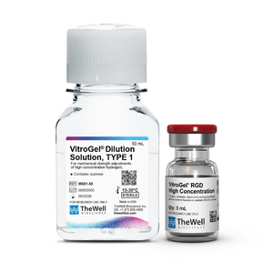
Hydrogels - Tunable
RGD modified - tunable, synthetic, chemically defined hydrogel - high concentration (3 mL kit)
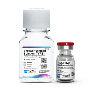
Hydrogels - Tunable
Laminin-derived functional peptide (IKVAV) modified – tunable, synthetic, chemically defined hydrogel, high concentration (3 mL kit)
Hydrogels - Tunable
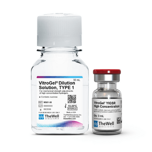
Laminin-derived functional peptide (YIGSR) modified – tunable, synthetic, chemically defined hydrogel, high concentration (3 mL kit)
Hydrogels - Tunable
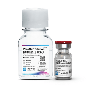
collagen-mimetic functional hydrogel - tunable, synthetic, chemically defined (3 mL)
Hydrogels - Tunable
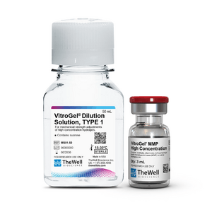
Matrix metalloproteinases (MMP) sensitive biodegradable hydrogel - tunable, synthetic, chemically defined hydrogel that supports biological activities such as cell proliferation, migration (adhesion/dispersion), differentiation, angiogenesis, apoptosis, etc.