APPLICATION NOTE
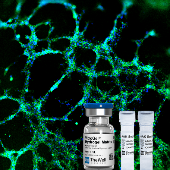
VitroGel® Angiogenesis Assay HC Kit
Tunable hydrogel system for 2D gel coating and 3D culture of angiogenesis tube formation, invasion, and animal injection.
VitroGel Angiogenesis Assay HC Kit
- Xeno-free functional hydrogel system supporting angiogenesis process
- Control and study the effect of the hydrogel mechanical properties on the angiogenesis process
- Multiple applications in one kit: Tube formation, invasion, and animal injection
- Control the hydrogel properties: Add your own growth factors and compare with positive control
- Harvest cells within 20 minutes at 37 °C without enzyme solution
VitroGel Angiogenesis Assay Kit is a revolutionary tool for researchers to study the effect of both hydrogel properties and culture medium on the angiogenesis process. The kit can be used to study the angiogenesis tube formation and invasion on both 2D hydrogel coating and 3D cell culture methods. The VitroGel system is also good for animal injection for in vivo studies.
Angiogenesis is a highly regulated process that involves the growth of new blood vessels from the existing vasculature. This process plays an important role in both normal developmental processes and numerous pathologies, including wound healing, tumor growth, and metastasis to inflammation and ocular disease.
Traditional angiogenesis assay highly relies on natural extracellular matrix (natural ECM), which has non-adjustable hydrogel compositions and properties. Therefore, our understanding of the angiogenesis process is limited by studying the molecular cues such as growth factors and inhibitors in culture medium only. There is a lack of knowledge on how hydrogel properties affect the angiogenesis process.
There are two versions of VitroGel Angiogenesis Assay Kits:
- VitroGel Angiogenesis Assay Kit (Cat No. VHM06-K): Ready-to-use with a fixed hydrogel mechanical strength to support the angiogenesis assay with adjustable supplements.
- VitroGel Angiogenesis Assay HC Kit (Cat No. TWG011-K): Assay kit with a tunable high-concentration hydrogel to allow full control of the hydrogel’s mechanical strength with adjustable supplements.
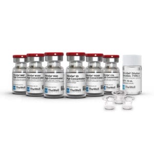
Depending on the kit type, the tunable VitroGel Angiogenesis Assay HC Kit can contain the following components:
- VitroGel AAK-HC, a tunable, xeno-free high-concentration hydrogel.
- AAK Dilution Solution, for adjusting the hydrogel concentration
- AAK Supplement 1, a hydrogel supplement without vascular endothelial growth factors (VEGFs) for promoting cell attachment and growth.
- AAK Supplement 2, a hydrogel supplement with VEGFs to promote tube formation and as a positive control.
All the components can be purchased separately.
Additional AAK Supplements can be purchased separately here >
Besides molecular cues, the VitroGel Angiogenesis Assay HC Kit allows researchers to explore the effects of hydrogel mechanical properties on angiogenesis. The high concentration VitroGel AAK-HC hydrogel is room temperature stable and can be adjusted by simply mixing the hydrogel solution and dilution solution at different rations (recommend 1:1 to 1:5 v/v) to achieve different mechanical strengths. The diluted hydrogel solution can be directly mixed with supplements at 2:1 (v/v) ratio for hydrogel formation. Researchers can adjust the molecular cues of the hydrogel by adding the growth factors/inhibitors directly to the supplement before mixing with VitroGel AAK-HC. Cells cultured in this system can be further harvested easily with the VitroGel Cell Recovery Solution.
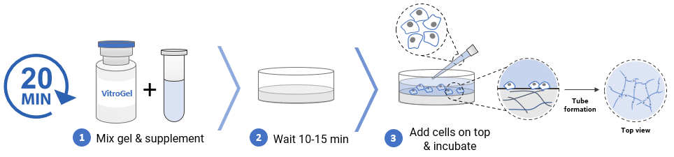
2D Hydrogel Coating for Tube Formation and Invasion Workflow
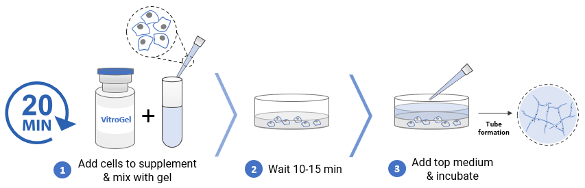
3D cell Culture Workflow
Specifications
| TYPE 1 Kit Contents | |
| TYPE 2 Kit Contents | |
| TYPE 3 Kit Contents | |
| Formulation | |
| Use | Angiogenesis Assay, tube formation, invasion, animal injection |
| Biocompatibility | Biocompatible, safe for animal studies |
| Injection | Injectable hydrogel for in vivo studies and lab automation |
| Cell Harvesting | VitroGel Organoid Recovery Solution 5-15 min cell recovery |
| pH | Neutral |
| Shipping | Supplements require dry ice shipment. |
| Storage | |
| Number of Uses | 30-180 tests per kit |
Recommended Product
Recover cells from the hydrogel in 20 minutes with high cell viability. Enzyme-free system.
Protocols / Handbooks / Resources
Product Documentations
![]() Product Data Sheet
Product Data Sheet![]() Frequently Asked Questions
Frequently Asked Questions![]() Material Safety Data Sheet (MSDS)
Material Safety Data Sheet (MSDS)
Video Protocols & Demonstrations
Application Notes
Data and References
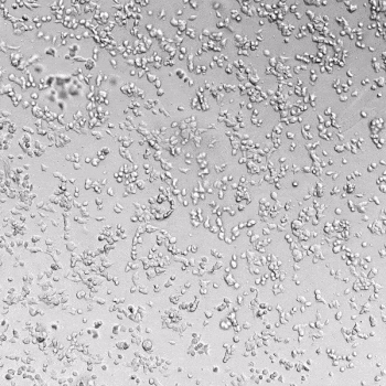
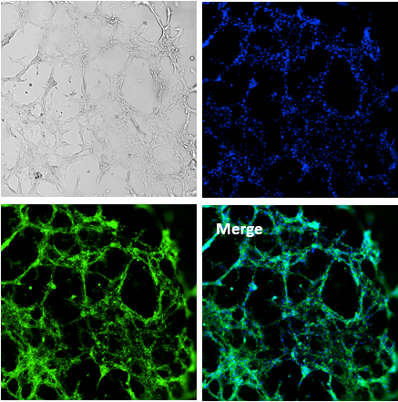
Figure 1. Tube formation of endothelial cells on top of VitroGel AAK-HC hydrogel with tube formation supplement, AAK Supplement 2.
The time-lapse video above shows the tube formation during 18 hours after Human Umbilical Vein Endothelial Cells (HUVEC) seeding on top of VitroGel AAK-HC hydrogel with AAK Supplement 2. The image above shows the tube morphology of HUVEC cells on top of VitroGel AAK-HC hydrogel. The cells were fixed and stained with DAPI (blue) and ActinGreen (green).
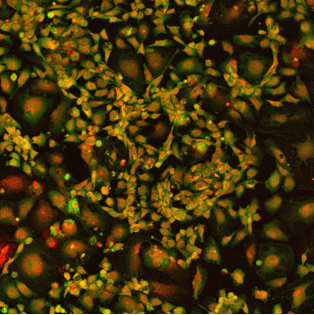
Figure 2. HUVEC cell growth on top of VitroGel AAK-HC hydrogel with cell growth supplement, AAK Supplement 1.
The image above shows HUVEC cells attached and grew on the surface of VitroGel AAK-HC hydrogel with cell growth supplement, AAK Supplement 1. The cells were fixed and stained with DRAQ5 (red) and actin green (green).
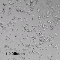
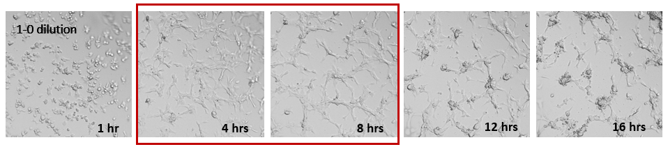
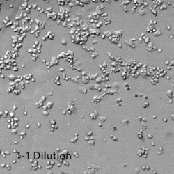
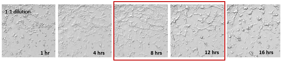
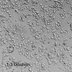
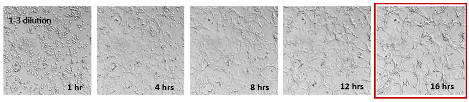
Figure 3. Effects of hydrogel mechanical strength on HUVEC tube formation.
Different concentrations of VitroGel AAK-HC hydrogels were prepared by diluting the high concentration VitroGel AAK-HC with AAK-HC Dilution Solution at 1:0, 1:1, and 1:3 (v/v) ratios. The hydrogels were then mixing with AAK Supplement 2 (with VEGFs for tube formation) and incubated in the 96-well plate for 15 minutes. HUVECs cell were added on top of the hydrogel and observed under a live cell imaging system. The time-lapse videos show the tube formation in 18 hours after cells added on top of hydrogel with different mechanical strength. The images indicate stronger hydrogel mechanical strength provided earlier cell tube structure formation (4-8 hours) than softer hydrogel (8-12 hours for 1:1 dilution and around 16 hours for 1:3 dilution). On the other hand, after tube formation, cells on the surface of 1:0 dilution hydrogel started to aggregate and form cell spheroids (around 12 hr). A similar aggregation happened to the cells on the surface of 1:1 diluted hydrogel (around 16 hr). For the soft hydrogel (1:3 dilution), the cells can attach well on the surface of the hydrogel and gradually form the tube structure within 18 hr. The results show the strong effect of the hydrogel mechanical strength on endothelial cell tube formation.
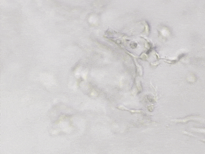
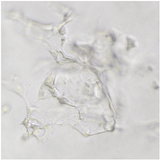
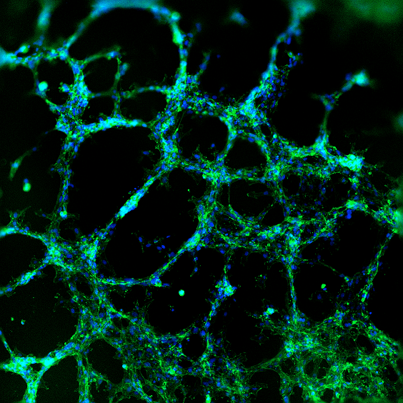
Figure 4. 3D tube structure of HUVEC cells cultured with the VitroGel Angiogenesis Assay HC Kit.
The concentration of VitroGel AAK-HC hydrogel was adjusted by diluting with AAK-HC Dilution Solution. HUVEC cells were prepared in hydrogel supplement before mixing with diluted hydrogel solution for 3D culture. The video shows the z-stack images of 3D tube structure formed inside of the hydrogel (after 7 days of incubation). The image above shows the tube networking structure inside of VitroGel AAK-HC hydrogel.
References/Publications
| AAK Kit | TYPE 1, TYPE 2, TYPE 3, Hydrogel Kit |
|---|
Related products
Assay Kits
A simple and easy replacement for animal-based ECM for consistent cell invasion studies. This kit includes the VitroGel® Hydrogel Matrix with VitroPrime™ Cell Culture Inserts, 8 µm, PET
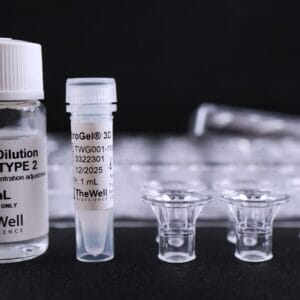
Xeno-free hydrogel system for consistent study of cell invasion. Kit includes choice of tunable functionalized hydrogel, 10mL VitroGel® Dilution Solution TYPE 2, and VitroPrime™ Cell Culture Inserts, 8 µm.
Angiogenesis Assay Kits
ready-to-use, hydrogel system for 2D gel coating and 3D culture of angiogenesis tube formation, invasion, and animal injection.
NEW
Ready-to-use kit for 3D GBM tumoroid formation. Supports epithelial-to-mesenchymal transition with long-term tumoroid culture. Hydrogel can be purchased separately. Click here