Research Highlights
Shining a (near-infrared) Light to Find Cancer
Institutions:
Shandong University and University of Jinan, China
Team:
Chen Y., Zhang X., Xiaoya L., Wu H., Zhang D., Zhu B., and Huang S.
Application:
HSC2 or CAL-27 cells in VitroGel xenografted into nude mice- identification of hypoxic levels in vivo
Disease Model:
Cancer progression and diagnosis
Hydrogel:
VitroGel® 3D Kit
Cancer cells grown in VitroGel 3D can be detected and tested for malignancy using a fluorescent chemical probe
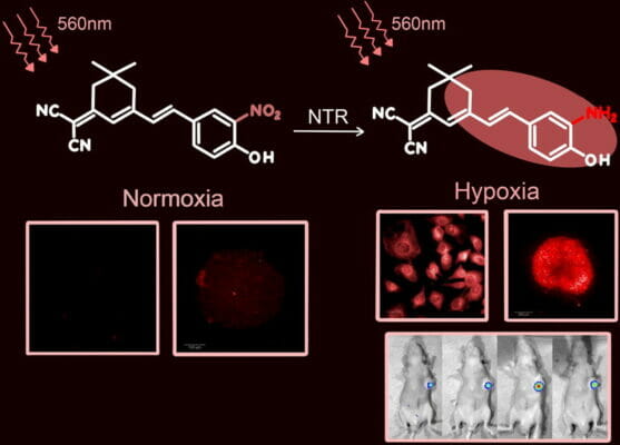
When certain cancer cells become malignant, they tend to cause a local reduction in the oxygen supply to the tissue. This state is called hypoxia. The lowered O2 concentration can helps tumors be more aggressive, invading nearby tissues during metastasis. It can also complicate anti-therapies by impeding the efficiency of radiation treatment and chemotherapy. However, hypoxia could also potentially be used to assist in our ability to detect and monitor tumor progression. It turns out that the enzyme nitroreductase (NTR) is overexpressed during conditions of hypoxia. This enzyme, if visualized with a chemical probe, then could be exploited as a means to track tumor location and growth. The aim of this paper was to develop a convenient fluorescent chemical probe for NTR that could be used to determine whether a tumor was benign or malignant.
A recent study out of the Cheeloo College of Medicine at Shandong University in China sought to synthesize and test a viable NTR fluorescent probe. The team was hopeful that a probe that signaled the presence of NTR and that could be detected using near-infrared (NIR) light could be found. Near-infrared light is advantageous because of its long wavelengths that penetrate deep into tissue and do not typically interact with molecules that are abundant in normal tissues, including nucleotides. Therefore, the team synthesized a molecule named HNT-NTR, which, when acted upon by the enzyme NTR, is converted into a product that has a characteristic emissions spectrum when irradiated with NIR light. To test the efficacy of HNT-NTR to characterize tumors, it was injected into mice that had been xenografted with two different types of tumor-forming cells. In order to prepare these cells (CAL-27 and HSC2 cells) for xenografting, they were first fixed in our VitroGel 3D hydrogel matrix. Approximately 1–10 million cells were mixed with 50 μL VitroGel 3D hydrogel and 50 μL of 0.5X PBS, and then injected into the armpits of BALB/c-nude mice. Tumors were seen after 2–3 weeks, and then these tumors had the HNT-NTR compound injected directly in them. Upon radiation with NIR light, the HNT-NTR (which had been shown to be non-toxic) displayed a distinct yellow-to-orange color change if present. And its presence in hypoxic cells, via conversion at the hands of NTR, was confirmed in the mice. Furthermore, when the mice were treated with anti-cancer agents such as cisplatin or 5-fluorouracil, a drop in the light emission from HNT-NTR injected cells could be seen, affirming the fact that this probe could be used to track tumor cells in vivo and provide a diagnostic reflection of their aggressiveness.
These experiments allowed the conclusion of three things. First, a chemical probe could be synthesized that is non-toxic, a substrate of nitroreductase, and responsive to probing with near-infrared light. Second, the probe can be safely and efficiently injected into tumors and persist in a mouse model. And third, the probe could allow detection and tracking of the state of hypoxia in a tumor, which in turn allows for an easy and accurate diagnosis of whether a tumor is likely to be benign or malignant.
Read the publication:
Related Products:
