Research Highlights
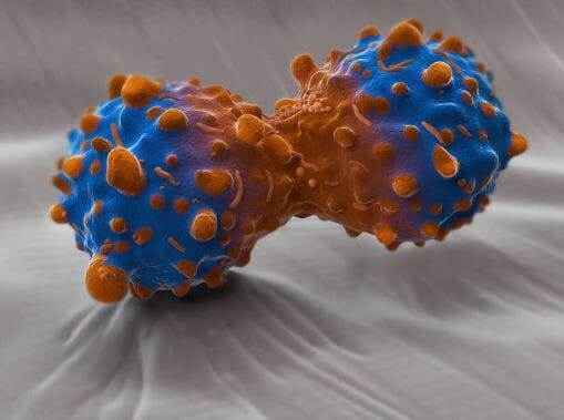
Breakdown in Communication: Extracellular Vesicles Drive Lung Cancer Progression by Shuttling c-Myc
Cancer cell-derived and plasma-derived oncogenic extracellular vesicles uniquely carry the cell cycle activator c-Myc and promote cell cycle upregulation and cluster formation of healthy cells in a 3D microenvironment.
Institution:
Fondazione IRCCS Istituto Nazionale dei Tumori, Istituto di scienze e tecnologie Molecolari CNR
Team:
Cristina Borzi, Linda Calzolari, Anna M. Ferretti, Laura Caleca, Ugo Pastorino, Gabriella Sozzi and Orazio Fortunato*
Application:
3D culture for proliferation assay
Disease model:
Lung Cancer
Cell types:
A549, LT73, BEC-KRASV12high cells
Hydrogel:
VitroGel® 3D (TWG001)
Lung cancer is one of the most common and malignant types of cancer and has maintained a high mortality rate because the early stages of this type of cancer do not exhibit obvious symptoms. Moreover, there has been a lack of effective therapeutic interventions because the molecular mechanisms underlying lung cancer development and progression remain poorly understood. It is known that lung tumorigenesis relies on a complex interplay between tumor and stromal cells and that these interactions are mediated by several mechanisms including cell-cell contact, paracrine signals. Recent studies have implicated spherical, bilayered, membranous vesicles known as extracellular vesicles (EVs) and can include exosomes, microvesicles, and apoptotic bodies. EVs are released by various cell types to facilitate cell-to-cell communication and do so by shuttling biomolecules to nearby or distant cells. Recent evidence has suggested that EVs play a crucial role in the growth and progression of lung cancer by modulating angiogenesis and epithelial-to-mesenchymal transition. What isn’t understood is how these tumorigenic extracellular vesicles contribute to lung tumorigenesis and what molecular players are involved in.
A recent study published in Cell Death and Disease by Cristina Borzi and colleagues sought to answer some of these questions. They used oncogenically modified bronchially epithelial cells to develop a model by which to demonstrate the communication between tumorigenic and non-tumorigenic cells and to understand how the cell-cell communication mediated by the EVs contributed to lung tumorigenesis. They found that EVs from the oncogenically transformed cells carried the cell cycle promoter c-Myc and that isolated EVs from tumorigenic cells had the ability to transform non-tumorigenic cells. Notably, they not only found c-Myc in extracellular vesicles isolated from cancer cells, but there were also able to detect it in EVs isolated from the blood plasma of lung cancer patients. It is also notable that c-Myc was not found in EVs isolated from healthy individuals having strong implications for the role of EVs in the cell-cell communication that leads to cancer metastasis.
One major disconnect between cancer research and the understanding of tumorigenesis and, therefore, the development of effective therapies has been the use of 2D culture to study an inherently 3D system. Significant limitations in 2D cancer research have included imprecise recapitulation of physiological cell-cell interactions, resulting in inaccuracies in gene expression. Moreover, considering the importance of the interaction of tumor cells with other stromal cells and the extracellular matrix during tumorigenesis, recapitulation of this phenotype is critical to understanding the molecular mechanism of tumorigenesis in vitro. To ensure the best possible model, Borzi and colleagues used The Well Biosciences VitroGel® 3D to test the 3D growth capacity and proliferation ability of cells exposed to EVs compared to non-tumorigenic cells. Using this ready-to-use tunable hydrogel, they were able to demonstrate that the EVs promoted tumorigenesis by enhancing proliferation, a finding in line with their discovery of c-Myc in the cancer-derived EVs. Their study laid down the foundation for ongoing mechanistic studies identifying the role of extracellular components like EVs in cancer malignancy, ultimately opening the door to improved understanding of lung cancer development and improved therapies.
Related Product:
