Application Notes
3D Invasion of Glioblastoma Cells in VitroGel® Hydrogel System
Application Note
Nana-Fatima Haruna, John Huang
TheWell Bioscience, North Brunswick, NJ 08902
Introduction
Glioblastoma is the most common primary tumor in the central nervous system. With an incidence of 3–5 out of 100,000 persons in Western countries, glioblastoma is one of the most deadly forms of malignant tumors, which has a median survival time of fewer than 15 months. The disease is characterized by poor prognosis as a result of the diffuse infiltrative growth pattern of the tumor cells into healthy brain tissue and spinal cord1,2. This pattern causes treatment failure, relapses, and poor survival rates. Therefore, the study of the signaling pathways, molecules, and processes that glioma cells modulate their surrounding environment is crucial in discovering novel treatments of glioblastoma3,4. Cell invasion is one of the highly mitotic activities of glioblastoma cells. Cells penetrate through an extracellular matrix (ECM) to perform a three dimensional (3D) migration. This is a very complex process that involves the changes in the migrating cell itself as well as the tumor microenvironment.
Besides tumor metastasis, cell invasion is also involved in a variety of physiological processes such as embryonic development, wound healing, immune responses, and angiogenesis. The mechanism of cell invasion involves strong cell-ECM interactions and the regulation of many biochemical and biophysical factors of the microenvironment5,6. For example, the invasion process in wound-healing is coordinated by different growth factors including platelet-derived growth factor (PDGF-BB), basic fibroblast growth factor (bFGF), and granulocytes macrophage colony-stimulating factor (GM-CSF)7. These growth factors stimulate cell types like keratinocytes, fibroblasts, endothelial cells, and macrophages to facilitate wound healing. Similarly, for cancer metastasis, cells interact with the ECM binding ligands to promote proliferation and the development of invadopodia-like structures.
Previously, the invasion assay studies relied on natural ECM such as Matrigel. These natural matrices contain a variety of biological components, including proteins, growth factors, and hormones in unknown concentrations, which make it hard to identify the crucial effects of specific factors on cellular behaviors. Moreover, it is hard to adjust the biochemical and mechanical properties of the natural matrices such as binding ligands and mechanical stiffness to fit different experimental designs. VitroGel®, a xeno-free and tunable hydrogel system, can solve these issues. The synthetic hydrogel system offers many advantages over native ECM hydrogels and allows scientists to study biological activity both in vitro and in vivo. In addition to increased consistency, VitroGel® hydrogels have been modified with functional ligands from fibronectin, collagen, laminin, and matrix metalloproteases (MMP) to create a well-defined system. Importantly, all these different functional modifications are offered in the same xeno-free tunable hydrogel 3D culture system, allowing scientists to investigate different combinations of VitroGel®products to find the right conditions for their experiments. This key feature permits the examination of cellular responses in a more physiologically relevant context, while still giving experimenters full control.
In this study, we used VitroGel® 3D and VitroGel® RGD to evaluate the invasion of glioblastoma cells (U-87 MG) in the horizontal direction. VitroGel® 3D is the pure hydrogel matrix without functional ligand modification and VitroGel® RGD is modified with integrin-binding ligand (RGD) to promote the cell adhesion and cell-matrix interactions. The mobility of U-87 MG cells in response to the integrin-binding effects of the hydrogel matrix can be examed by comparing cell movement in VitroGel® 3D and VitroGel® RGD. The unique shear-thinning properties of VitroGel® allow us to create a cell-free zone (hole) at the center of the hydrogel. By filling the hole with hydrogel contenting inducing factors (high concentration FBS in this case), we can make a concentration gradient within the hydrogel to induce cell migration. Also, the tunable VitroGel® system allows us to adjust the mechanical strength of the hydrogel by simply changing the hydrogel concentration. The cell movement into the void in response to the hydrogel’s mechanical property can be observed. This invasion model will also be useful in obtaining physiologically relevant data in wound healing, developmental biology, regenerative medicine, and cancer studies.
Materials and Methods
Cell Culture
Human glioma cells (U-87 MG) were maintained in Alpha Minimum Essential Medium supplemented with 10% Fetal Bovine Serum (FBS). The cells were passaged when the cultures reached 80-90% confluence. The cells were trypsinized and suspended in complete cell culture medium with 10% FBS at a concentration of 1-2 x 10⁶ cells/mL.
Prepare hydrogel for 3D Horizontal Invasion of U-87 MG cells
The 3D invasion of U-87 MG cells was tested in VitroGel® through the following preparation steps:
- Dilute hydrogel solution of VitroGel® 3D and VitroGel® RGD with VitroGel® Dilution Solution (Type 2) at 1:3 and 1:5 (v/v) ratios according to the user handbook.
- Mix the diluted hydrogel solution and the cell suspension at 4:1 (v/v) ratio.
- Add 50 μL of the hydrogel-cell mixture to each well in a 96 well plate. The final concentration of FBS in the hydrogel was 2%.
- The hydrogel was left to stabilize for 30-60 minutes in a 5% CO2 incubator at 37°C.
- Prepare the second hydrogel mixture with a high concentration of FBS without cells: use diluted hydrogel solution from step 1 to mix with 100% FBS at 4:1 (v/v) ratio to get the hydrogel mixture with 20% FBS.
- After stabilization of step 4, a micropipette was used to create a 5-10 μL cell-free detection zone by sucking the hydrogel mixture out (as shown in the diagram below).
- The zone was immediately filled with the hydrogel mixture from step 5 to create a concentration gradient of FBS within the hydrogel.

Rheology
The rheological properties of the 1:3 and 1:5 diluted hydrogel were examined to match the mechanical properties of the hydrogel to the cell response observed in both conditions. To test the rheological properties of the stable hydrogel, the samples were prepared in a petri dish; 30min later, an additional medium was added to the gel surface and the plate was incubated at 37°C overnight to allow for hydrogel solidification. The tests were performed using a Malvern Kinexus dynamic rheometer (Malvern Panalytical, UK) at a 0.1% controlled shear rate model.
Cell Imaging
The cells were stained with calcein-AM to produce green-fluorescence in live cells. Projection images of the cells embedded in the hydrogel were created with different Z levels and taken at 4X and 10X magnification using ImageXpress (Molecular Devices, CA) on days 1 and 7 to show the horizontal movement of the cells into detection. The stronger cell attachment and cell-matrix interactions in RGD modified hydrogel promoting cell mobility is consistent with previous reports that glioma cells are sensitive to the integrin-binding ligands4,8. In addition to the FBS gradient attracting cells, the adhesive ligand plays an essential role in supporting cell invasion within the ECM.
Results
Figure 1 shows the cell invasion over time in VitroGel® hydrogels. On day 0, there is a clear cell-free zone (detection zone) at the center of the hydrogel matrix. The high FBS concentration in the detection zone generated the attraction to induce cell movement into the zone. U-87 MG cell invasion is integrin-binding ligands dependent as shown by the different cell movement patterns and speed on VitroGel® 3D and VitroGel® RGD from day 1 to day 7 (figure 1). Only a small amount of cells moved into the detection zone after 7-day incubation in VitroGel® 3D. However, the number of cells in the detection zone was significantly increased in VitroGel® RGD after 7 days.
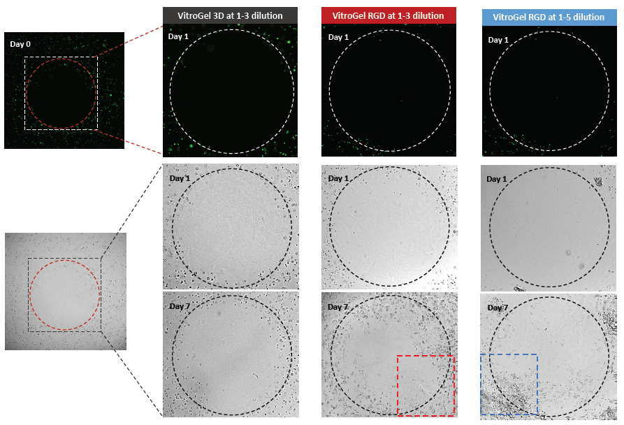
Figure 1: 3D invasion of U-87 MG cells in VitroGel® 3D (1:3 dilution) and VitroGel® RGD (1:3 and 1:5 dilution). The clear cell-free detection zone was observed on Day 0. The 3D migration of cells toward the high FBS concentration detection zone was observed by comparing the image data of day 1 and day 7. The images were taken at 4x magnification.
On the other hand, the ECM’s mechanical property could be another vital factor influencing cell mobility. Figure 1 shows the successful cell invasion in both 1:3 and 1:5 (v/v) diluted RGD hydrogel. However, when we look into more details (red and blue square of Figure 1), the enlarged images in figure 2 indicates that the cell movement is faster in the 1:5 (v/v) diluted hydrogel by showing more extending cells in the center area. The 1:3 hydrogel (400 Pa) supports the growth of the cells but from the result, the cell-matrix interaction is enhanced by the lower mechanical strength of the 1:5 hydrogel (100 Pa), allowing for the development and extension of epithelial structures and the formation of more complex cellular networks.
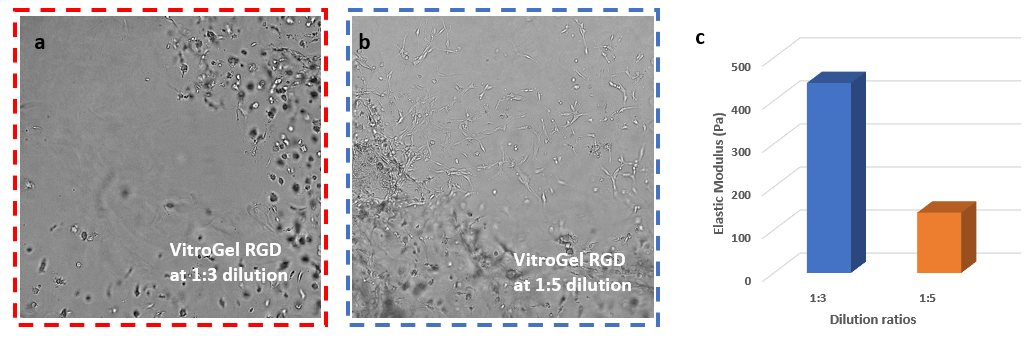
Figure 2: A portion of the detection zone in VitroGel® RGD 1:3 (a) and 1:5 (b) dilution hydrogels. The images were taken at 10x magnification on day 7. Cells in the 1:5 diluted hydrogel showed more epithelial projections and cell networking than the cells in the 1:3 diluted hydrogel. The final gel strength (c) of the 1:3 and 1:5 dilution hydrogels was measured at ≈400 Pa and ≈100 Pa respectively.
Discussion
In this study, the tunable hydrogel VitroGel® was used to set up a horizontal invasion assay through an extracellular matrix. The system supported cell growth, cell-matrix, and cell-cell interactions. This invasion model can study important biological responses, including migration, invasion, proliferation, and angiogenesis9,10.
The complex pathological and physiological processes involved in wound healing, inflammation, embryonic development, cancer, and cell differentiation can be studied with this 3D horizontal invasion model7,11,12. The cells used in these studies employ diverse mechanisms that involve complex interactions with their environment, such as the utilization of matrix metalloproteases (MMP) to digest and navigate the ECM during both metastasis and wound healing. Therefore, physiologically and clinically relevant cell-based assays for these studies need to be in tunable systems to account for the diverse interactions within the matrix14-15. The system also allows adhesive binding ligands, growth factors, and various other factors initiating complex cell behaviors to be studied and analyzed.
As the result shows, VitroGel® hydrogel can be effectively used for 3D cell exclusion zone assays for compound screening. The system enables the controlled combination of a variety of materials and biological properties, including MMP and other bio-functional ligands, to study cell behavior in more complex ways. It also provides a platform for a detailed and reproducible study of cell invasion under different mechanical and biochemical conditions.
Reference
- Stock, K. et al. Capturing tumor complexity in vitro: Comparative analysis of 2D and 3D tumor models for drug discovery. Scientific Reports 6, (2016).
- Riedl, A. et al. Comparison of cancer cells in 2D vs 3D culture reveals differences in AKT–mTOR–S6K signaling and drug responses. Journal of Cell Science 130, 203–218 (2016).
- Amann, A. et al. Development of a 3D angiogenesis model to study tumour – endothelial cell interactions and the effects of anti-angiogenic drugs. Scientific Reports 7, (2017).
- Wu, M. & Swartz, M. A. Modeling Tumor Microenvironments In Vitro. Journal of Biomechanical Engineering 136, 0210111–0210117 (2014).
- Brekhman, V. & Neufeld, G. A novel asymmetric 3D in-vitro assay for the study of tumor cell invasion. BMC Cancer 9, 415 (2009).
- Edmondson, R., Broglie, J. J., Adcock, A. F. & Yang, L. Three-Dimensional Cell Culture Systems and Their Applications in Drug Discovery and Cell-Based Biosensors. ASSAY and Drug Development Technologies 12, 207–218 (2014).
- Hulkower, K. I. & Herber, R. L. Cell Migration and Invasion Assays as Tools for Drug Discovery. Pharmaceutics 3, 107–124 (2011).
- Yu, X. & Machesky, L. M. Cells Assemble Invadopodia-Like Structures and Invade into Matrigel in a Matrix Metalloprotease Dependent Manner in the Circular Invasion Assay. PLoS ONE 7, e30605 (2012).
- Gnanamony, M. et al. Chronic radiation exposure of neuroblastoma cells reduces nMYC copy number. Oncology Letters 14, 3363–3370 (2017).
- Vinci, M., Box, C. & Eccles, S. A. Three-Dimensional (3D) Tumor Spheroid Invasion Assay. Journal of Visualized Experiments (2015). doi:10.3791/52686
- Antoni, D., Burckel, H., Josset, E. & Noel, G. Three-Dimensional Cell Culture: A Breakthrough in Vivo. International Journal of Molecular Sciences 16, 5517–5527 (2015).
- Melissaridou, S. et al. The effect of 2D and 3D cell cultures on treatment response, EMT profile and stem cell features in head and neck cancer. Cancer Cell International 19, (2019).
- Ceylan, H. et al. 3D-Printed Biodegradable Microswimmer for Theranostic Cargo Delivery and Release. ACS Nano 13, 3353–3362 (2019).
- Bellis, S. L. Advantages of RGD peptides for directing cell association with biomaterials. Biomaterials 32, 4205–4210 (2011).
- Baker, B. M. & Chen, C. S. Deconstructing the third dimension – how 3D culture microenvironments alter cellular cues. Journal of Cell Science 125, 3015–3024 (2012).