Why Study Cell Invasion?
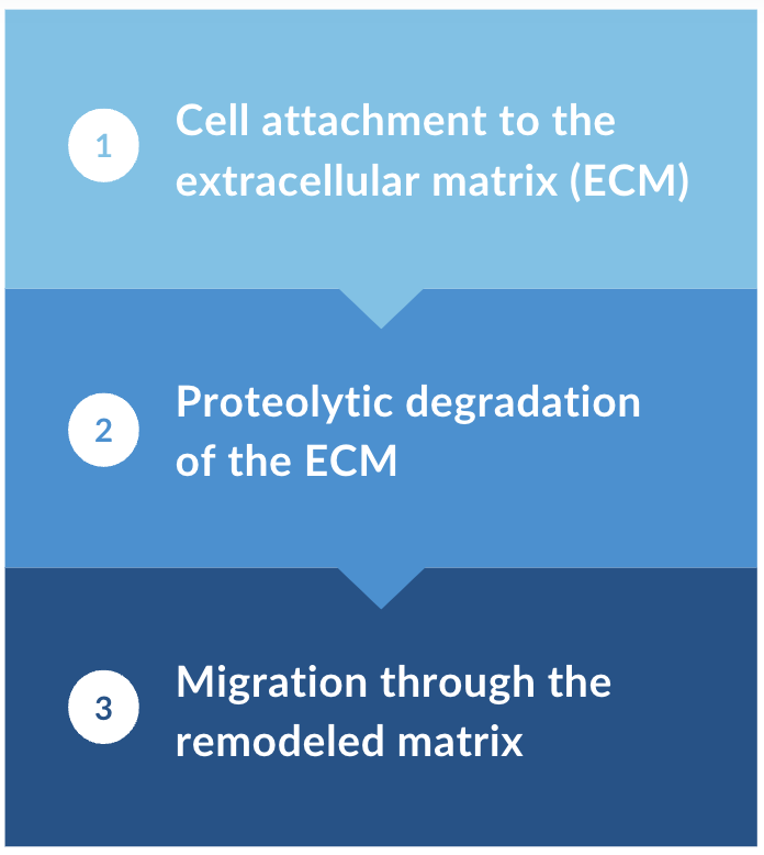
Cell invasion and migration are crucial for processes such as tissue development, immune responses, wound healing, and cancer metastasis. During invasion, cells undergo morphological changes, degrade the extracellular matrix, and respond to environmental signals.
Invasion assays are crucial for studying these processes, providing insights into normal functions and diseases. Understanding cell invasion is key for developing targeted therapies and improving cancer treatment outcomes.
General Steps for
Cell Invasion
Related Invasion Models
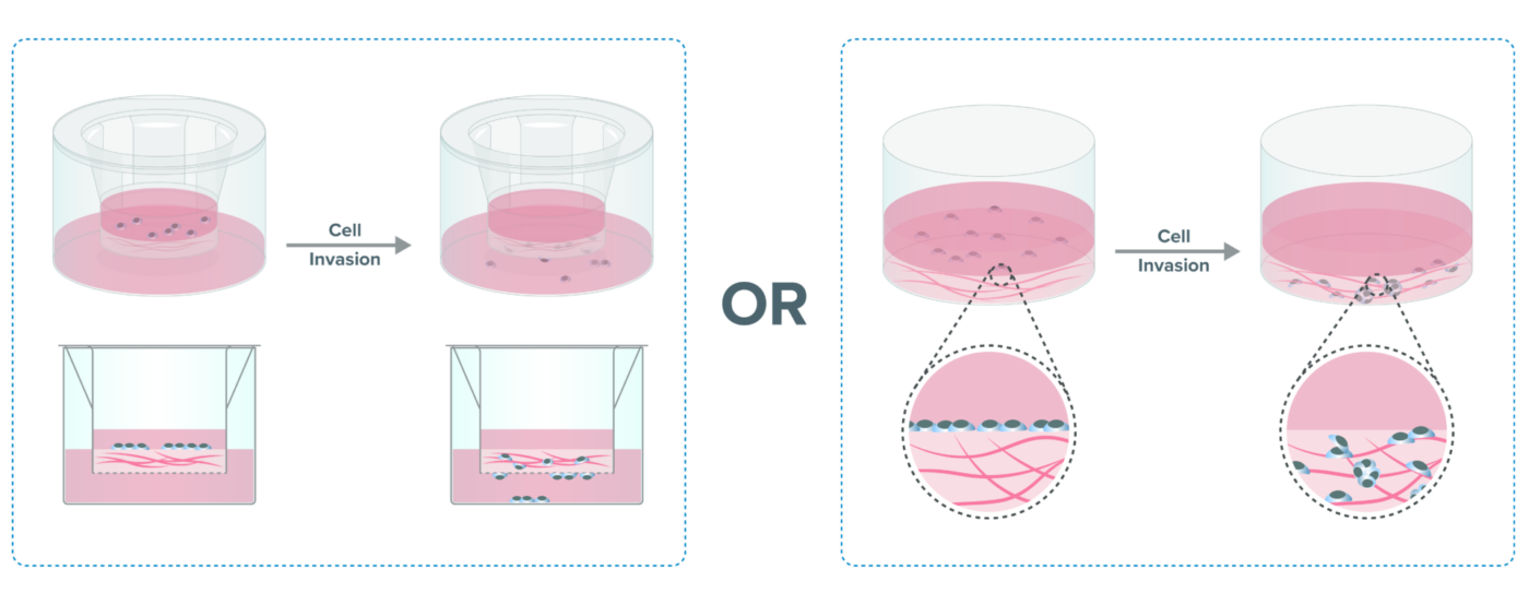
Barrier Model
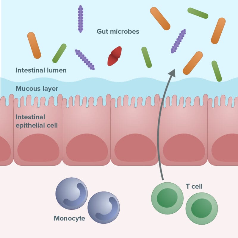
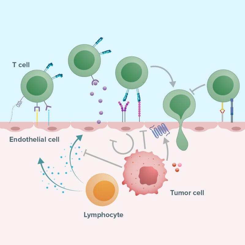
Immune Cell Infiltration
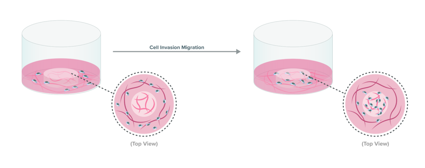
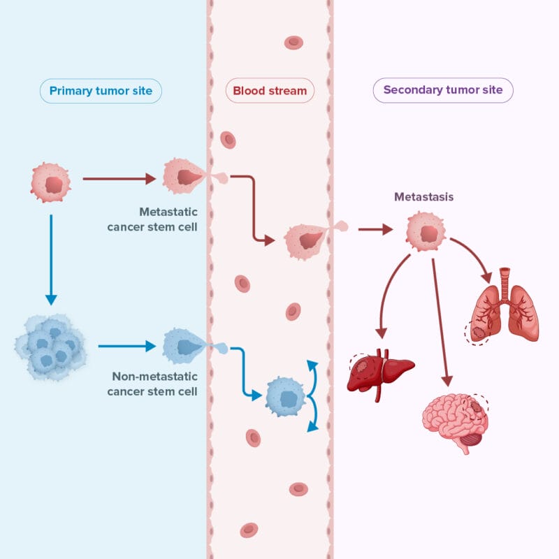
Cancer Metastasis
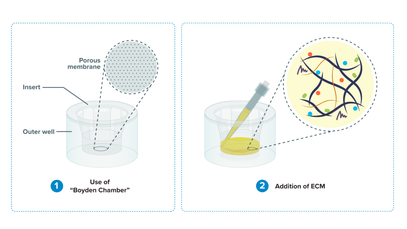
The role of ECM in cell invasion
Traditional cell invasion assay is performed in a “Boyden Chamber” system. A layer of extracellular matrix (ECM) is added on top of the membrane insert to create a barrier between the cells on top of the ECM and the medium in the outer chamber.
However, the traditional animal-based ECM contains complicated components that not only introduce unpredictability to cell invasion study but also cause a lack of understanding of the effect of ECM on cell mobility.
The extracellular matrix (ECM) mimics the in vivo environment, allowing:
- cells to interact with a natural scaffold
- influence cell adhesion and migration
- signaling pathways essential for invasion
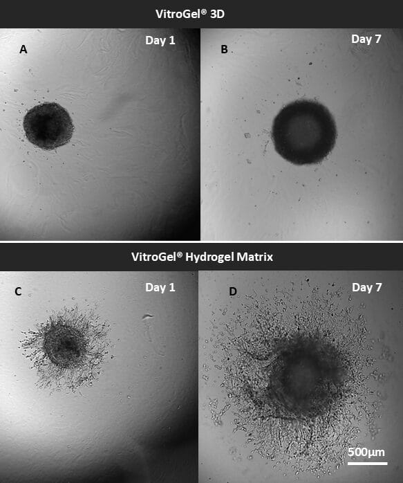
VitroGel®: A new tool to understand cell invasion
Our advanced VitroGel® hydrogel system closely replicates the native ECM’s physical and functional properties, offering a perfect balance of biological complexity and ease of use. Besides our ready-to-use hydrogel, our unique tunable hydrogel system allows for a detailed study of the microenvironment’s influence on cell mobility by adjusting factors like:

Mechanical Strength
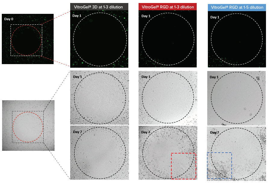
Study cell mobility under different mechanical strengths ranging from 10 to 4000 Pa. Just adjust the dilution ratio!.

Degradation
Study the effect of biodegradation on cell mobility using VitroGel® MMP.

Functional Ligand
Prepare your own multi-functional matrix taking advantage of “Mix and Match” capability of VitroGel® High-Concentration Hydrogels.

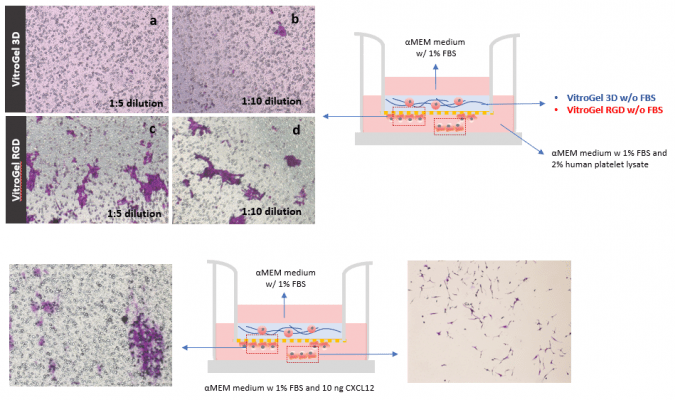
Serum/Growth Factors/ Cytokine/Chemokines
The different concentrations of serum, growth factors, and chemokines can be directly mixed with VitroGel® or added to the outside of the insert well to induce the cell movement.
VitroGel®
Hydrogels
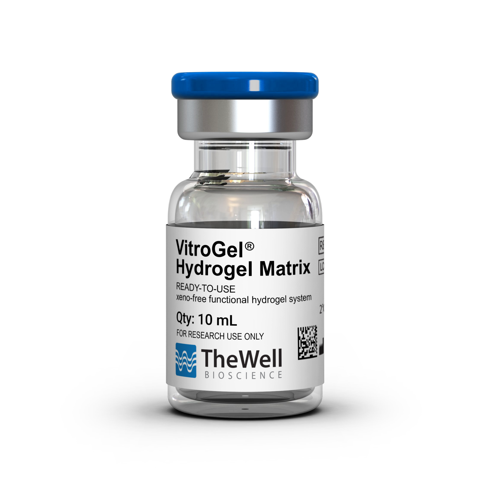
VitroGel® is a xeno-free (animal origin-free), versatile, biofunctional hydrogel that closely mimics the physiological extracellular matrix.
VitroGel®
Cell Invasion Assay Kit
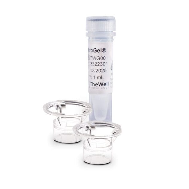
VitroGel® Cell Invasion Assay Kit includes a ready-to-use hydrogel and premium cell culture inserts/plate. A simple and easy replacement for animal-based ECM for consistent cell invasion studies.
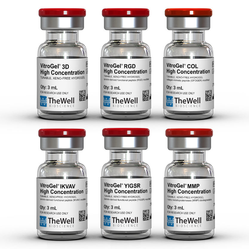
VitroGel® High-Concentration
Cell Invasion Assay Kit
VitroGel® High-Concentration Cell Invasion Assay Kit features a tunable, xeno-free hydrogel with premium inserts and plates, enabling customizable matrix stiffness, ligands, and ECM components.
CytoGrow™
Growth Factors

CytoGrow™ is TheWell Bioscience’s portfolio of premium-grade recombinant proteins built for researchers who demand top-tier bioactivity, strong lot-to-lot consistency, and reliable performance—with a research-friendly price.
VitroPrime™
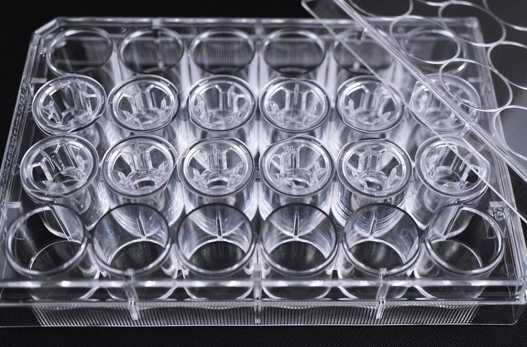
Cell Culture Inserts
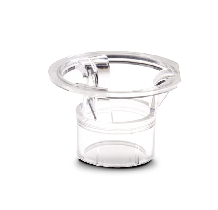
VitroPrime™ Cell Culture Inserts are premium quality cell culture inserts for consistent invasion studies. Excellent alternative to Transwell.
VitroPrime™ Ultra-Low
Attachment Plate

VitroPrime™ Ultra-Low Attachment,
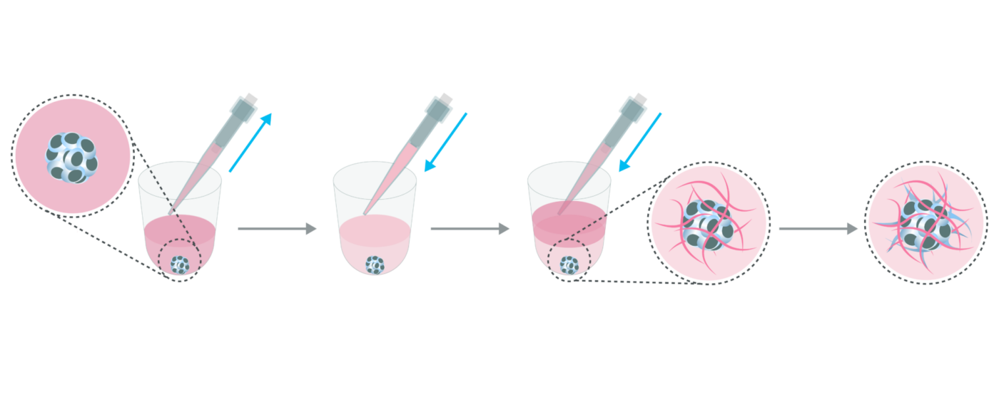
U-Bottom, 96-Well Plate is a premium plate that enables 3D spheroid formation and growth.
How to select VitroGel® for cell invasion assay
The simplest answer to this question is to choose the Ready-to-Use VitroGel® Cell Invasion Assay Kits (Cat #IA-VHM01-1P). This multifunctional hydrogel system has been designed to easily assay cell invasion and migration with a wide range of cell types.
For researchers who want to have an in-depth understanding of the relationship between the microenvironment and cell behaviors, the High-Concentration VitroGel® Cell Invasion Assay Kits, hydrogels provide an excellent system that can be easily manipulated in hydrogel strength, binding ligand, degradability, and supplement compositions.
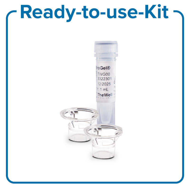
VitroGel® Cell Invasion Assay Kit
Components:
- VitroGel® Hydrogel Matrix
- VitroPrime™ Cell Culture Inserts
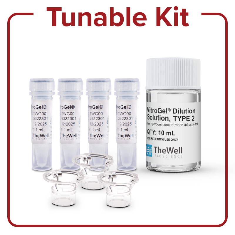
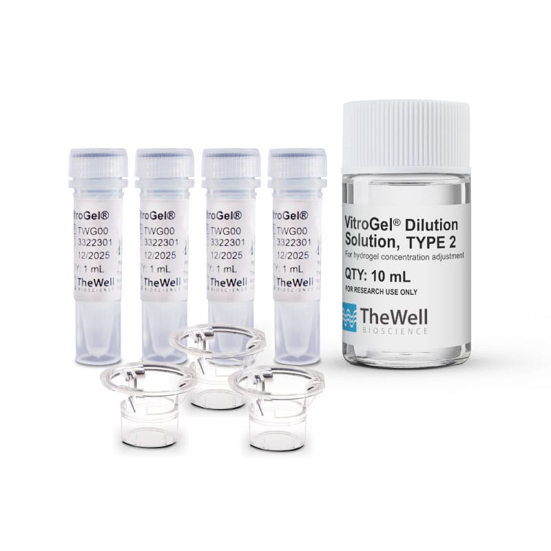
VitroGel® High-Concentration
Cell Invasion Assay Kit
Components:
- VitroGel® High-Concentration
- VitroGel® Dilution Solution
- VitroPrime™ Cell Culture Inserts